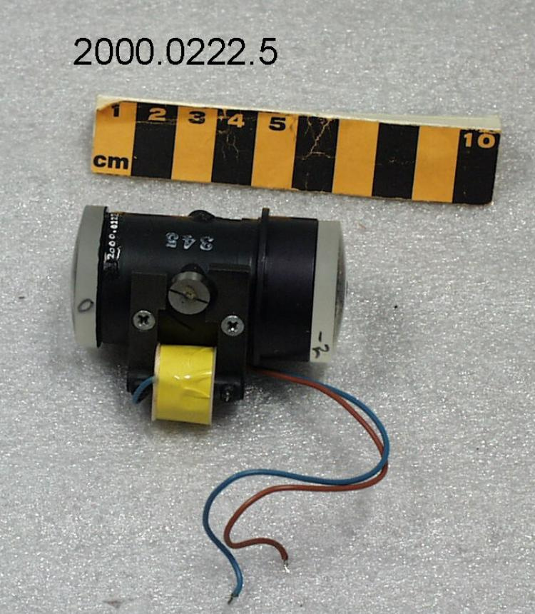
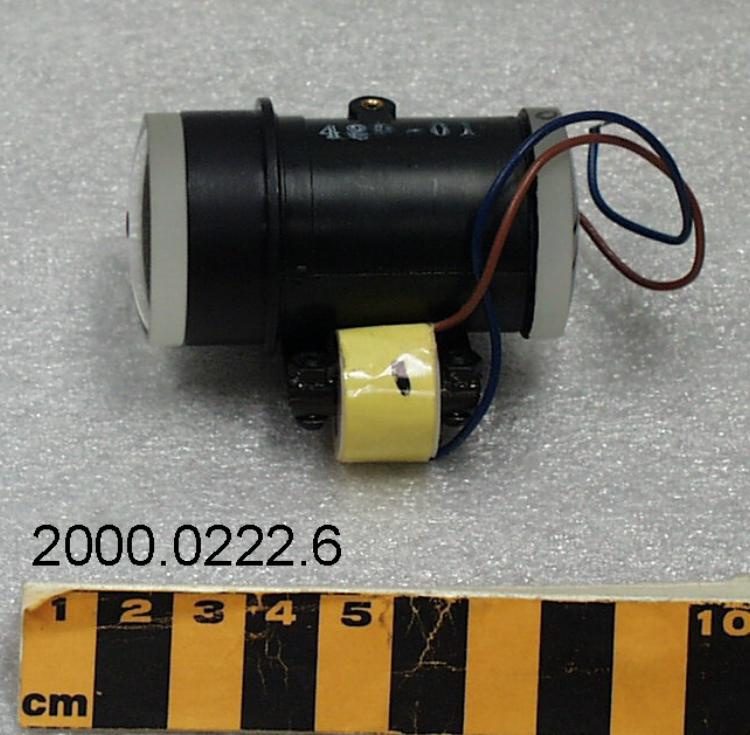
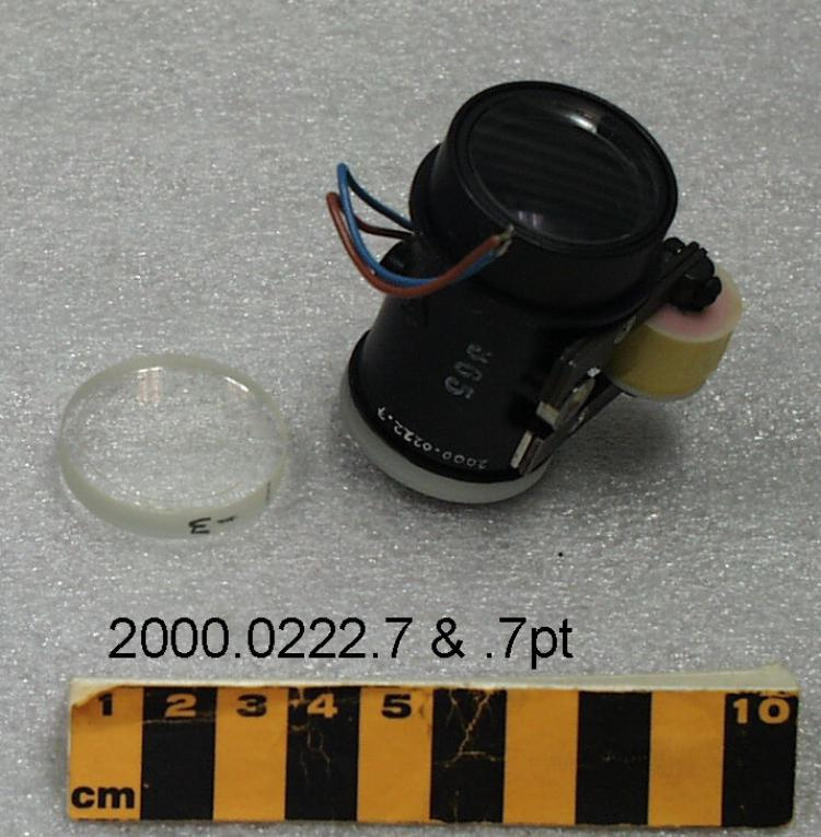
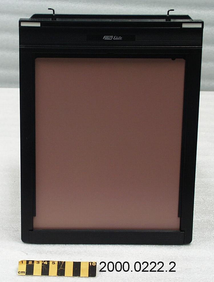
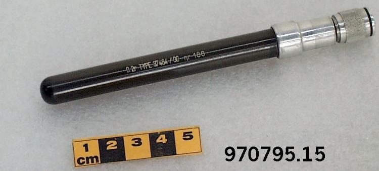
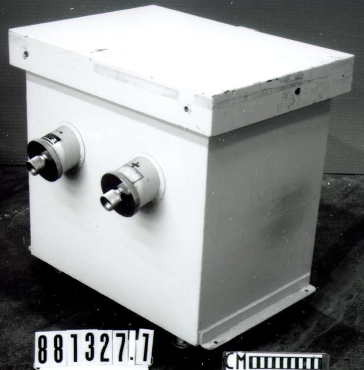
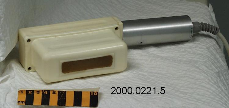
Transducer
Use this image
Can I reuse this image without permission? Yes
Object images on the Ingenium Collection’s portal have the following Creative Commons license:
Copyright Ingenium / CC BY-NC-ND (Attribution-NonCommercial 4.0 International (CC BY-NC 4.0)
ATTRIBUTE THIS IMAGE
Ingenium,
2000.0221.005
Permalink:
Ingenium is releasing this image under the Creative Commons licensing framework, and encourages downloading and reuse for non-commercial purposes. Please acknowledge Ingenium and cite the artifact number.
DOWNLOAD IMAGEPURCHASE THIS IMAGE
This image is free for non-commercial use.
For commercial use, please consult our Reproduction Fees and contact us to purchase the image.
- OBJECT TYPE
- hand held
- DATE
- 1987
- ARTIFACT NUMBER
- 2000.0221.005
- MANUFACTURER
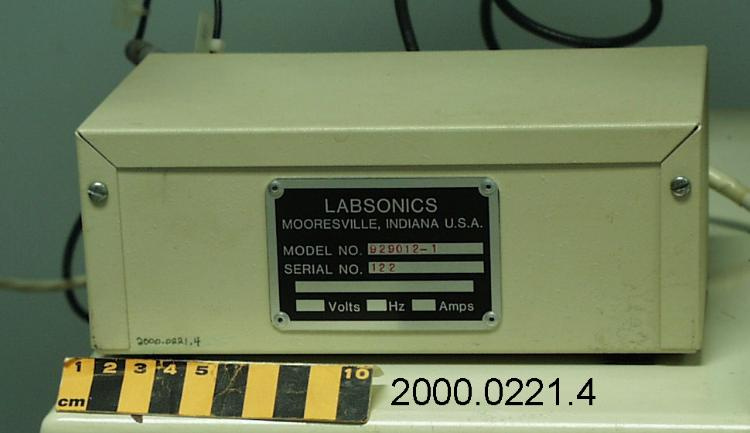
- LABSONICS INC.
- MODEL
- Unknown
- LOCATION
- Mooresville, Indiana, United States of America
More Information
General Information
- Serial #
- N/A
- Part Number
- 5
- Total Parts
- 10
- AKA
- wand
- Patents
- N/A
- General Description
- METAL, GLASS & SYNTHETIC.
Dimensions
Note: These reflect the general size for storage and are not necessarily representative of the object's true dimensions.
- Length
- 25.5 cm
- Width
- 5.3 cm
- Height
- 10.0 cm
- Thickness
- N/A
- Weight
- N/A
- Diameter
- N/A
- Volume
- N/A
Lexicon
- Group
- Medical Technology
- Category
- Radiology
- Sub-Category
- N/A
Manufacturer
- AKA
- LABSONICS
- Country
- United States of America
- State/Province
- Indiana
- City
- Mooresville
Context
- Country
- Canada
- State/Province
- Quebec
- Period
- THIS ULTRASOUND UNIT USED LATE 1980S- C. 2000.
- Canada
-
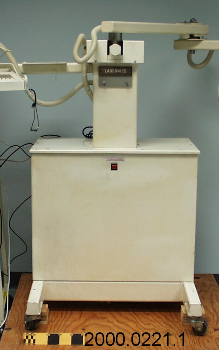
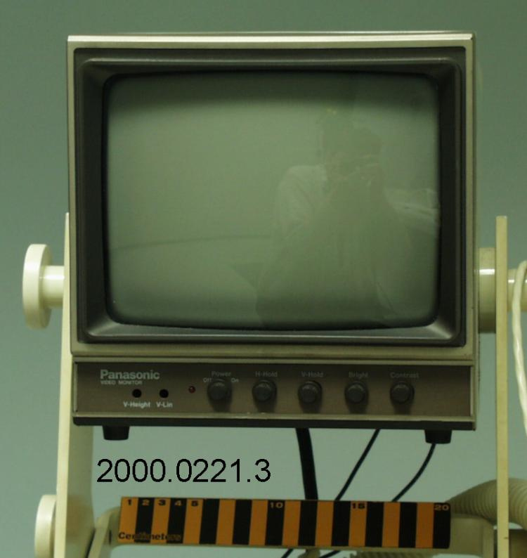
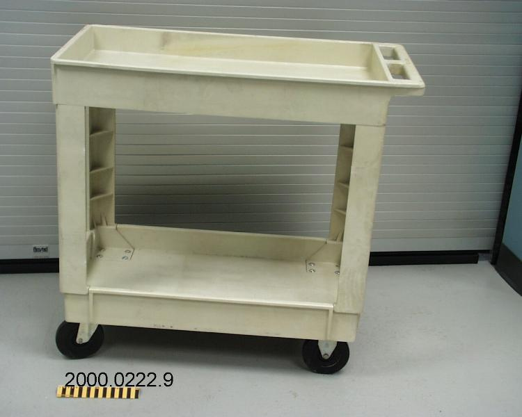
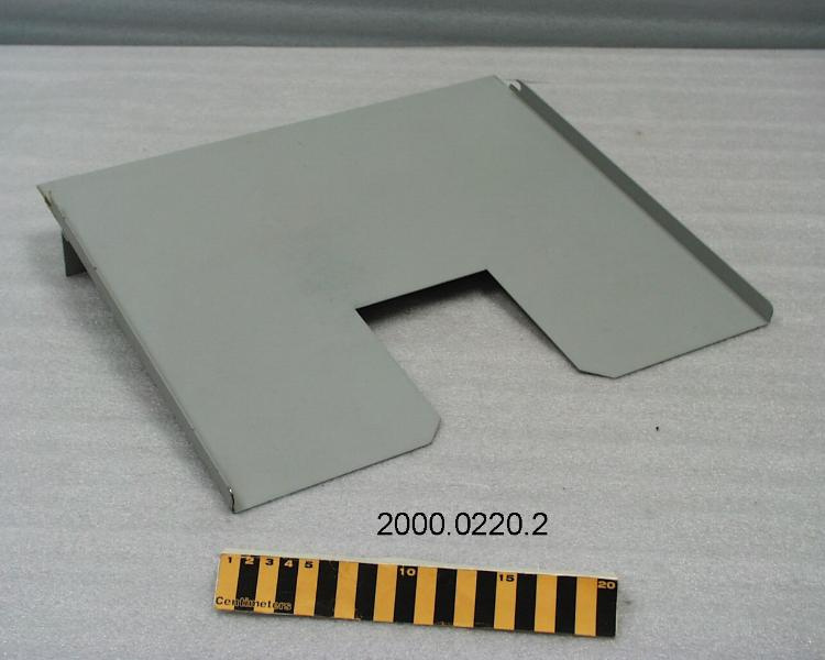
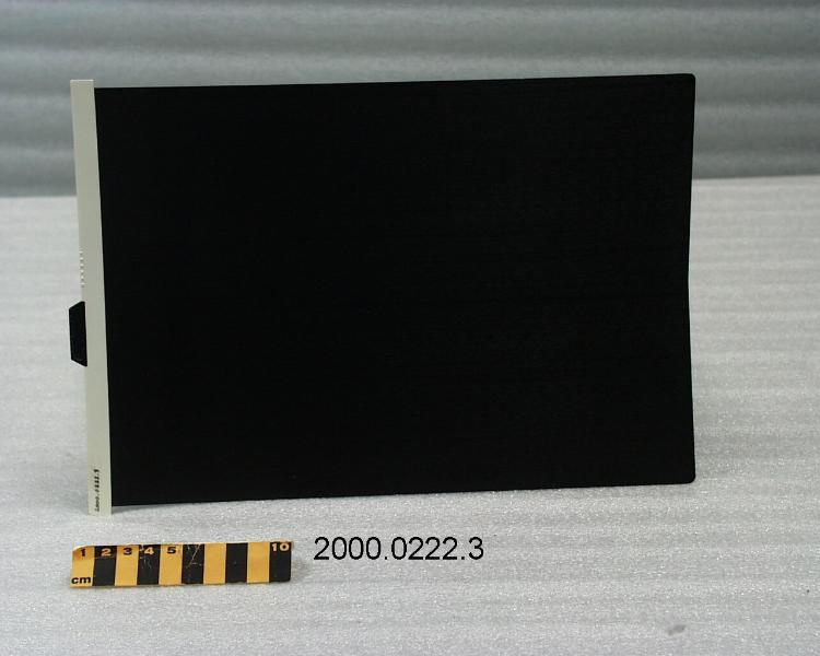
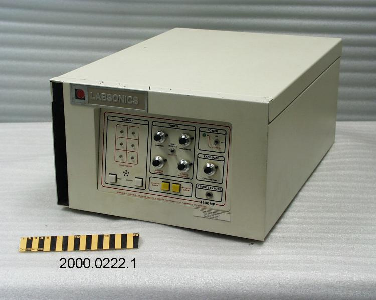
USED BY DR. ROGER GHYS IN HIS MONTREAL, PQ MEDICAL PRACTICE. DR. GHYS IS CONSIDERED TO BE AMONG THE FIRST CANADIAN PHYSICIANS TO ROUTINELY USE ULTRASOUND AND RELATED TECHNIQUES TO DIAGNOSE BREAST CANCER (LATE 1980S). DR. GHYS ALSO PROVIDED SURGEONS WITH POSITIONAL DATA (AND IMAGES) ACCURATE ENOUGH THAT MANY IMAGING PROCEDURES DID NOT HAVE TO BE REPEATED PRIOR TO SURGERY. THIS IS 1ST ULTRASOUND MACHINE USED IN CANADA FOR DIAGNOSTIC BREAST SCANS. (REF.1) - Function
-
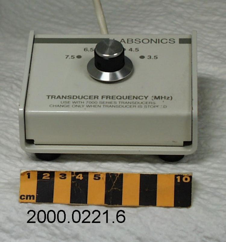
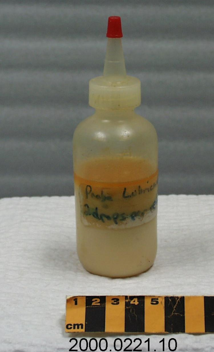
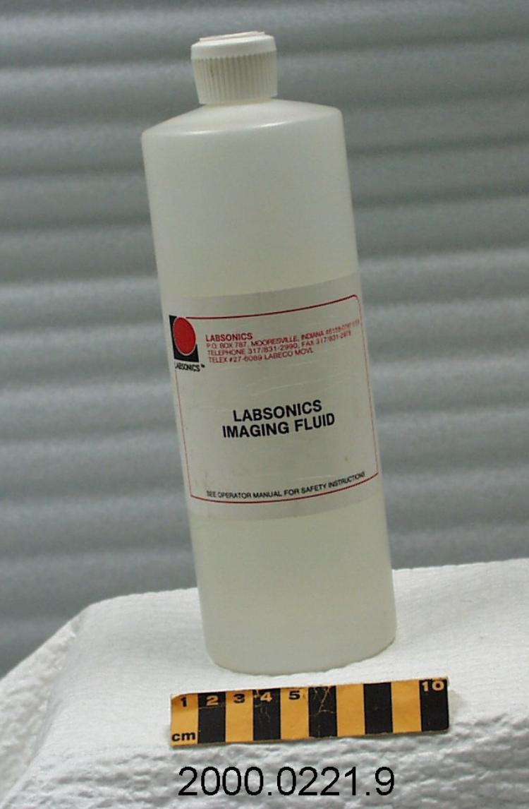
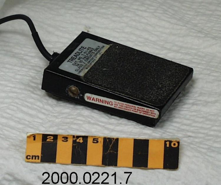
USED TO OBTAIN IMAGES FROM INSIDE THE BODY, IN ORDER TO OBSERVE MOVEMENT OF INTERNAL ORGANS AND TISSUES, BLOOD FLOW, THE PRESENCE OF SWELLING OR UNUSUAL GROWTHS, ETC. THIS UNIT USED TO OBSERVE CHANGES OR ABNORMALITIES IN BREAST TISSUE. - Technical
-
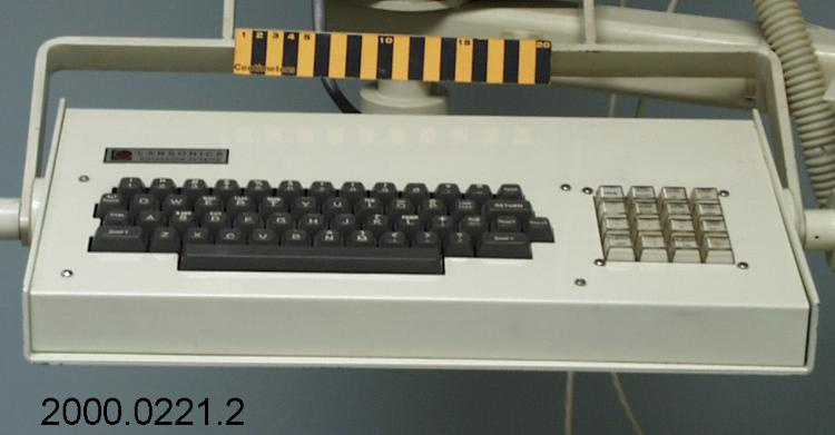
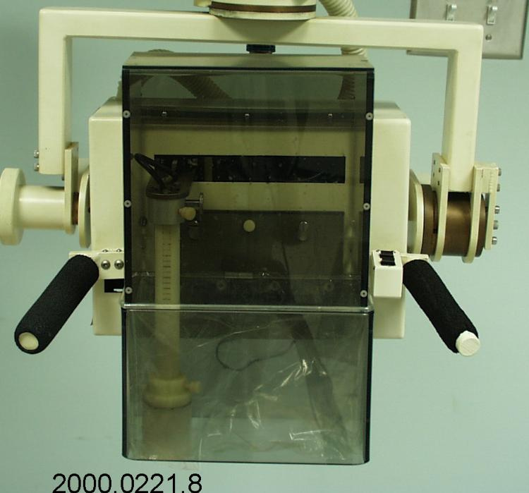
PROTOTYPE DESIGN ULTRASOUND UNIT MFD. BY LABSONICS WAS FAR ADVANCED OVER EXISTING ULTRASOUND APPARATUS. SQUARE IMAGE PRODUCED WAS LESS DISTORTED & ALLOWED PHYSICIAN TO MEASURE 3-D LOCATION OF POSSIBLE TUMORS WITH GREATER ACCURACY. SYSTEM COMBINES COMPUTER CONTROL AND ANALYSIS WITH THE CONTROL & DIGITAL CREATION OF THE IMAGE FOLLOWING THE CONCEPTS FIRST USED IN CAT SCANS. (REF. 1) - Area Notes
-
Unknown
Details
- Markings
- No markings visible.
- Missing
- N/A
- Finish
- COMPONENT HAS CREAM-COLOUR FINISH: BRUSHED SILVER METAL, GREY AND/OR BLACK TRIM.
- Decoration
- N/A
CITE THIS OBJECT
If you choose to share our information about this collection object, please cite:
LABSONICS INC., Transducer, circa 1987, Artifact no. 2000.0221, Ingenium – Canada’s Museums of Science and Innovation, http://collection.ingeniumcanada.org/en/id/2000.0221.005/
FEEDBACK
Submit a question or comment about this artifact.
More Like This
