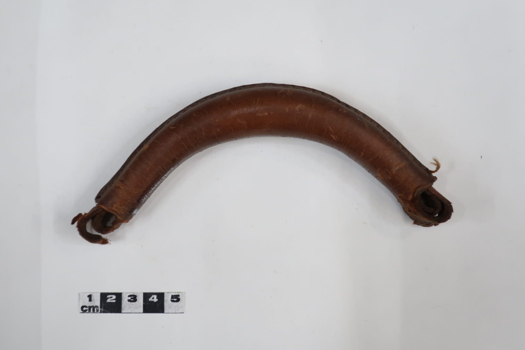
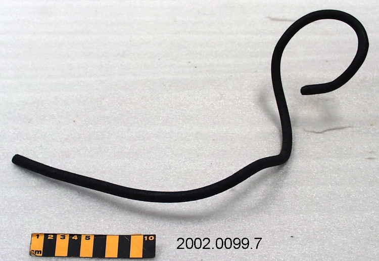
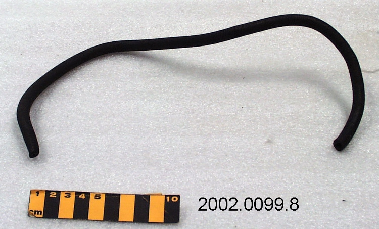
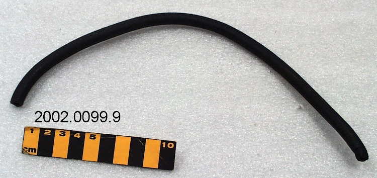
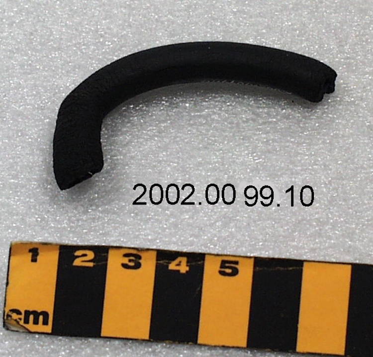
Tuyau
Utiliser cette image
Puis-je réutiliser cette image sans autorisation? Oui
Les images sur le portail de la collection d’Ingenium ont la licence Creative Commons suivante :
Copyright Ingenium / CC BY-NC-ND (Attribution-NonCommercial 4.0 International (CC BY-NC 4.0)
ATTRIBUER CETTE IMAGE
Ingenium,
1969.1029.009
Permalien:
Ingenium diffuse cette image sous le cadre de licence Creative Commons et encourage son téléchargement et sa réutilisation à des fins non commerciales. Veuillez mentionner Ingenium et citer le numéro de l’artefact.
TÉLÉCHARGER L’IMAGEACHETER CETTE IMAGE
Cette image peut être utilisée gratuitement pour des fins non commerciales.
Pour un usage commercial, veuillez consulter nos frais de reproduction et communiquer avec nous pour acheter l’image.
- TYPE D’OBJET
- S/O
- DATE
- 1925–1935
- NUMÉRO DE L’ARTEFACT
- 1969.1029.009
- FABRICANT
- General Electric X-ray Corp.
- MODÈLE
- Inconnu
- EMPLACEMENT
- Chicago, Illinois, United States of America
Plus d’information
Renseignements généraux
- Nº de série
- S/O
- Nº de partie
- 9
- Nombre total de parties
- 38
- Ou
- S/O
- Brevets
- S/O
- Description générale
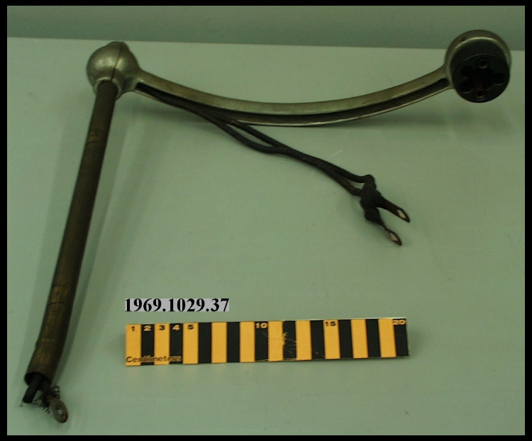
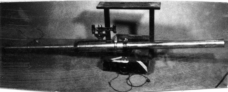
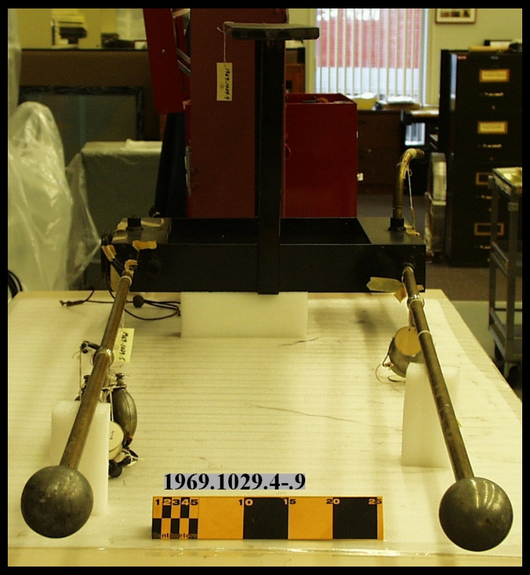
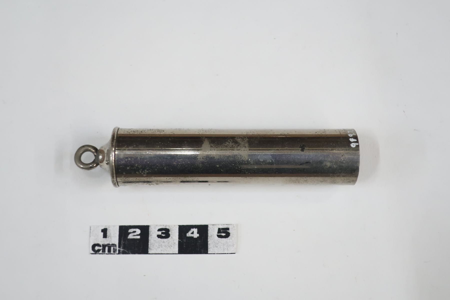
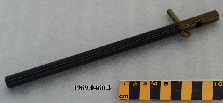
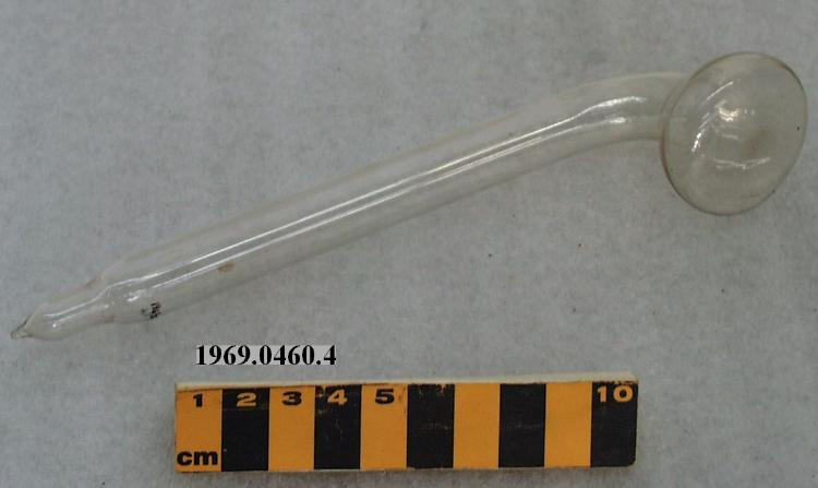
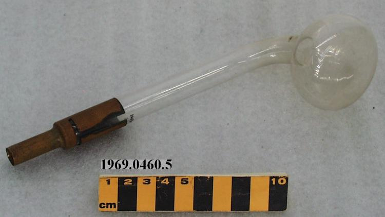
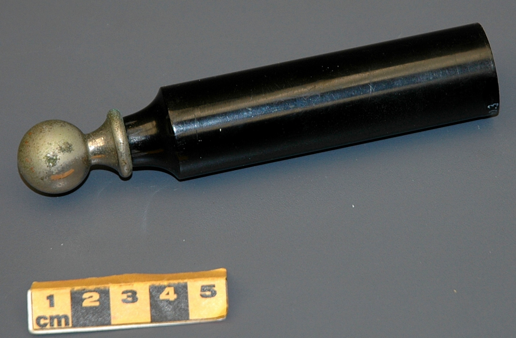
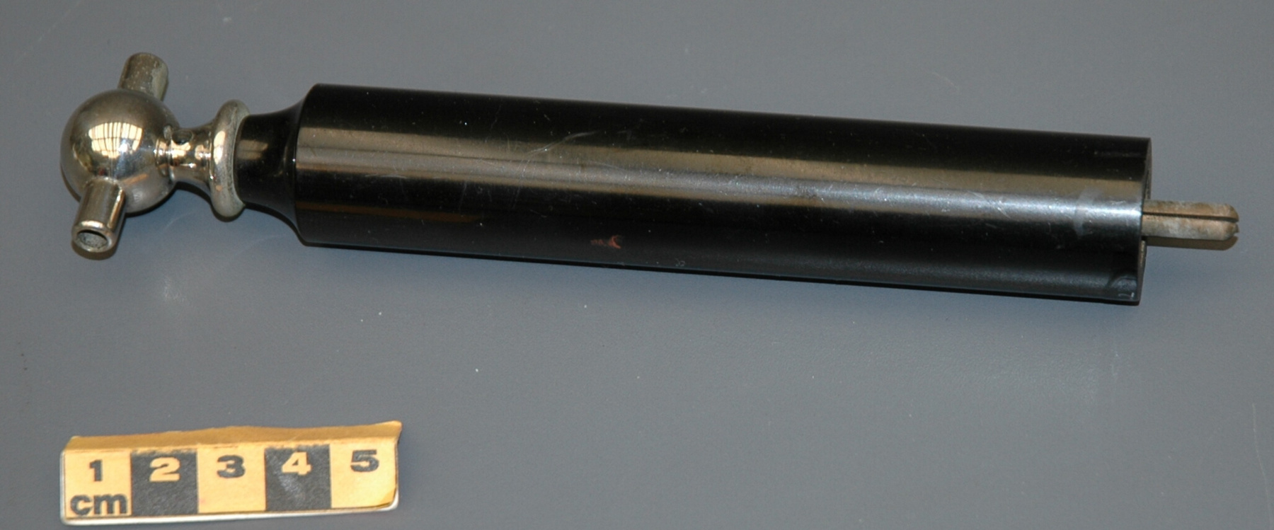
- Copper pipe
Dimensions
Remarque : Cette information reflète la taille générale pour l’entreposage et ne représente pas nécessairement les véritables dimensions de l’objet.
- Longueur
- 15,0 cm
- Largeur
- 14,0 cm
- Hauteur
- S/O
- Épaisseur
- S/O
- Poids
- S/O
- Diamètre
- 2,0 cm
- Volume
- S/O
Lexique
- Groupe
- Technologie médicale
- Catégorie
- Radiologie
- Sous-catégorie
- S/O
Fabricant
- Ou
- General Electric
- Pays
- United States of America
- État/province
- Illinois
- Ville
- Chicago
Contexte
- Pays
- Amérique du Nord
- État/province
- Inconnu
- Période
- This type of machine probably used c. late 1920s+
- Canada
-
Dr. Richard Proctor, University of Manitoba Graduate who served in France starting in 1915. Previously in charge of the X-Ray department in the Strathcona Hospital in Edmonton he is noted to have changed his Meyers X-Ray machine to a Victor in 1925. - Fonction
-
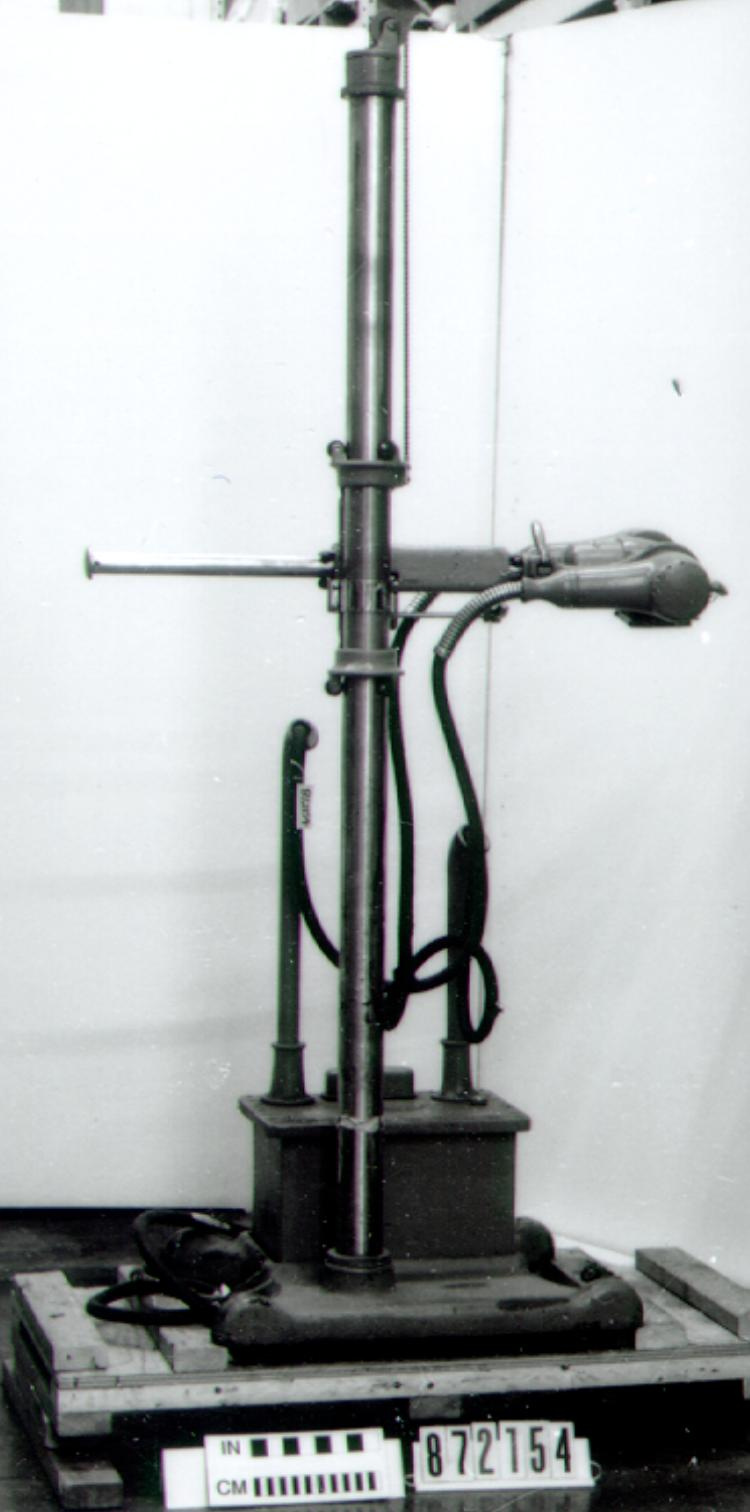
Used to produce graphic images of internal structures.S. Therapeutic X-ray sets exist for the treatment of skin and superficial disorders, and these consist of an X-ray generator and suitable positioning and beam collimating equipment. However, the majority of X-ray apparatus is for the production of images on film or video screens for diagnostic purposes. 6 - Technique
-
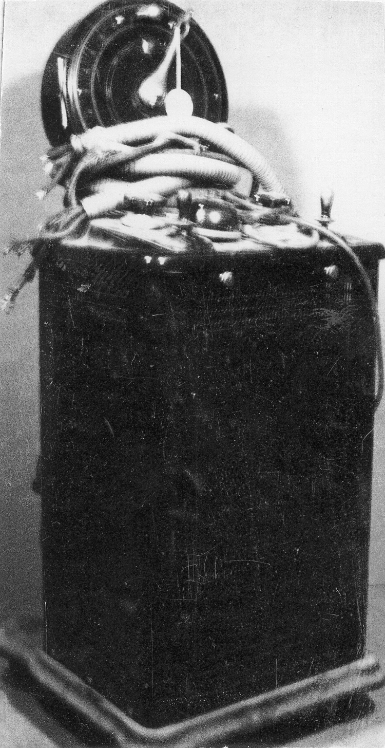
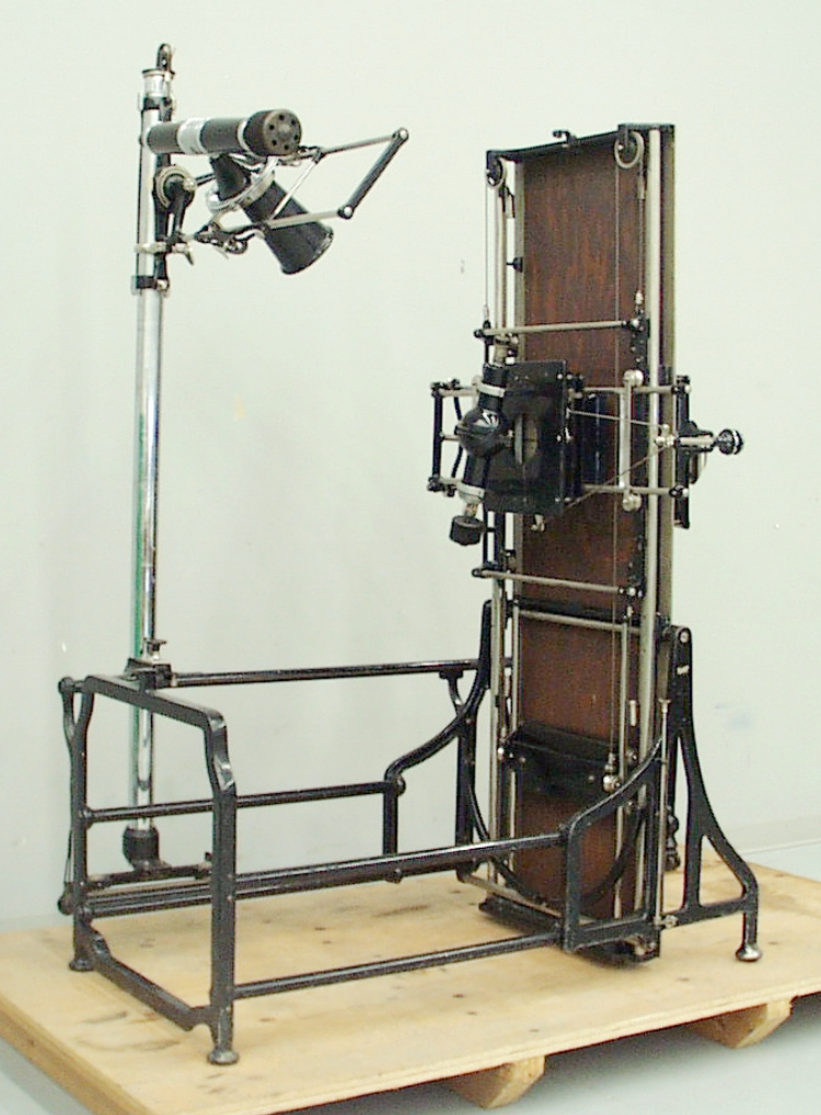
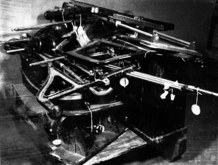
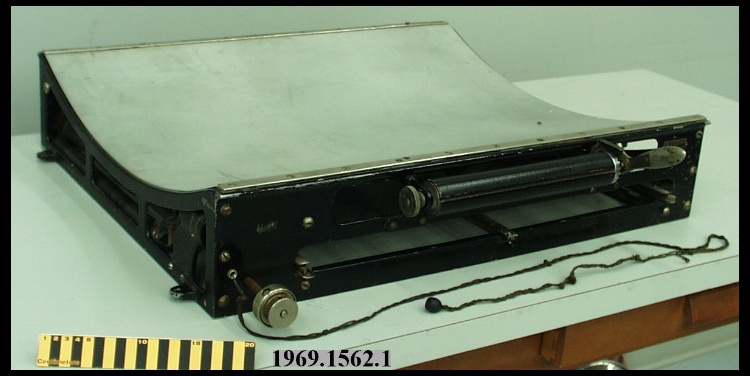
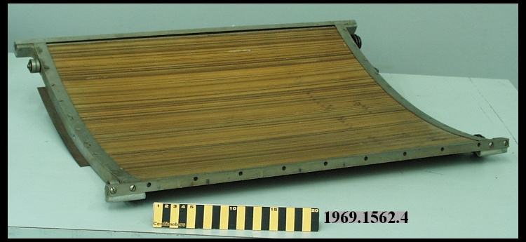
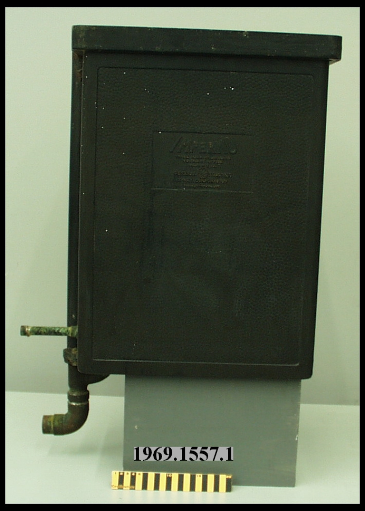
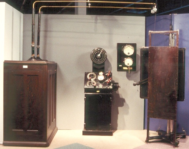
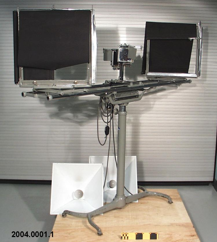
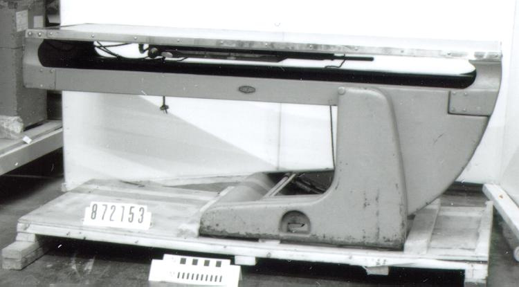
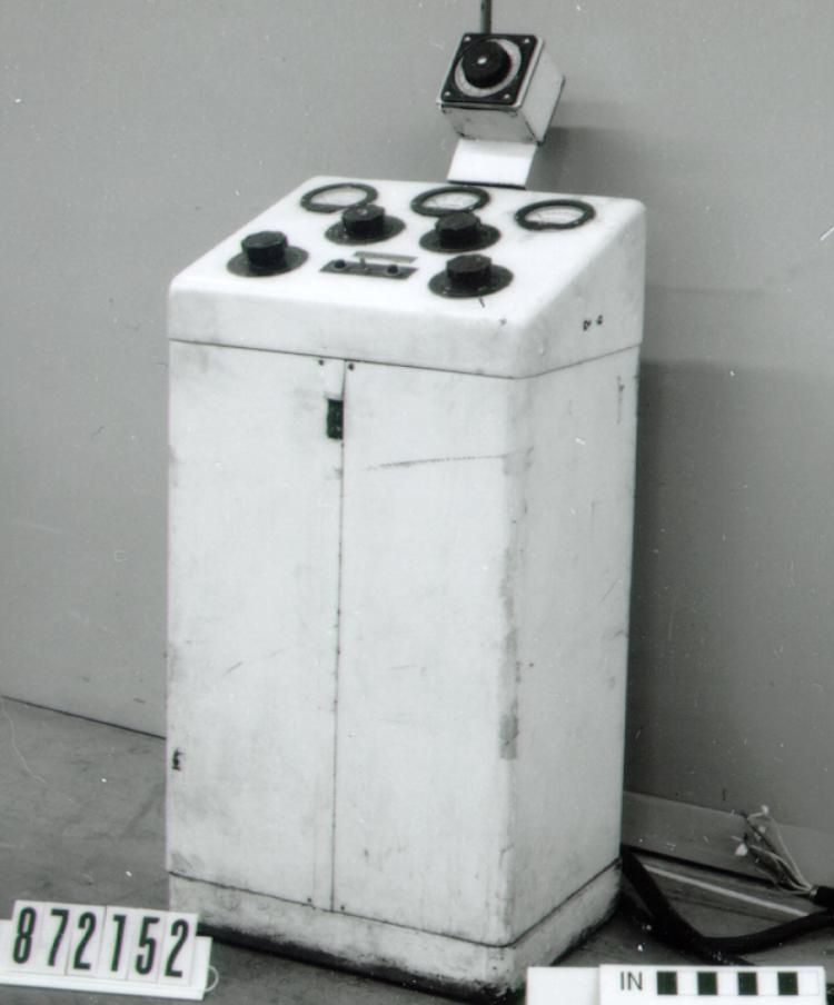
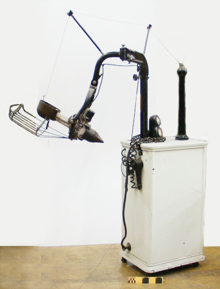
Early, elaborate x-ray machine features both fluoroscoping (diagnostic) and radiographic (treatment) apparatus, and a table capiable of both vertical and horizontal positioning. Probably used with separate curved potter-bucky. X-ray machines were able to take only single films. The unit usually has the tube mounted above, and the film below. The tube can usually be moved to various positions and angles including the horizontal position for chest films, in which case the cassette and Bucky are in a separate stand. 6 - Notes sur la région
-
Inconnu
Détails
- Marques
- "G" handwritten in blue ink on masking tape label fixed to pipe.
- Manque
- S/O
- Fini
- Curved length of copper pipe.
- Décoration
- S/O
FAIRE RÉFÉRENCE À CET OBJET
Si vous souhaitez publier de l’information sur cet objet de collection, veuillez indiquer ce qui suit :
General Electric X-ray Corp., Tuyau, vers 1925–1935, Numéro de l'artefact 1969.1029, Ingenium - Musées des sciences et de l'innovation du Canada, http://collection.ingeniumcanada.org/fr/item/1969.1029.009/
RÉTROACTION
Envoyer une question ou un commentaire sur cet artefact.
Plus comme ceci


