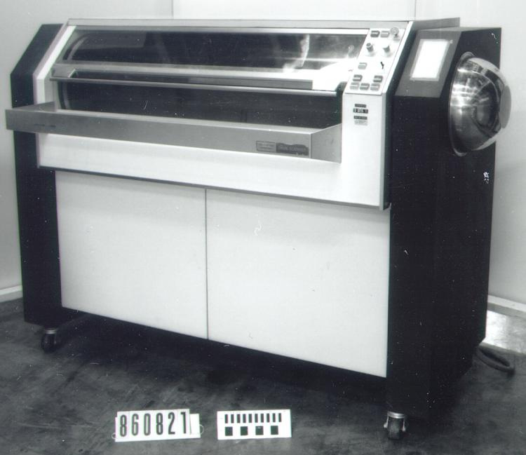
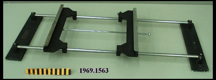
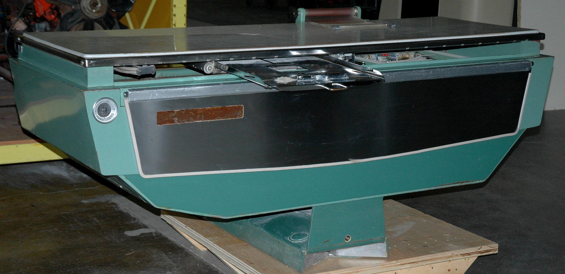
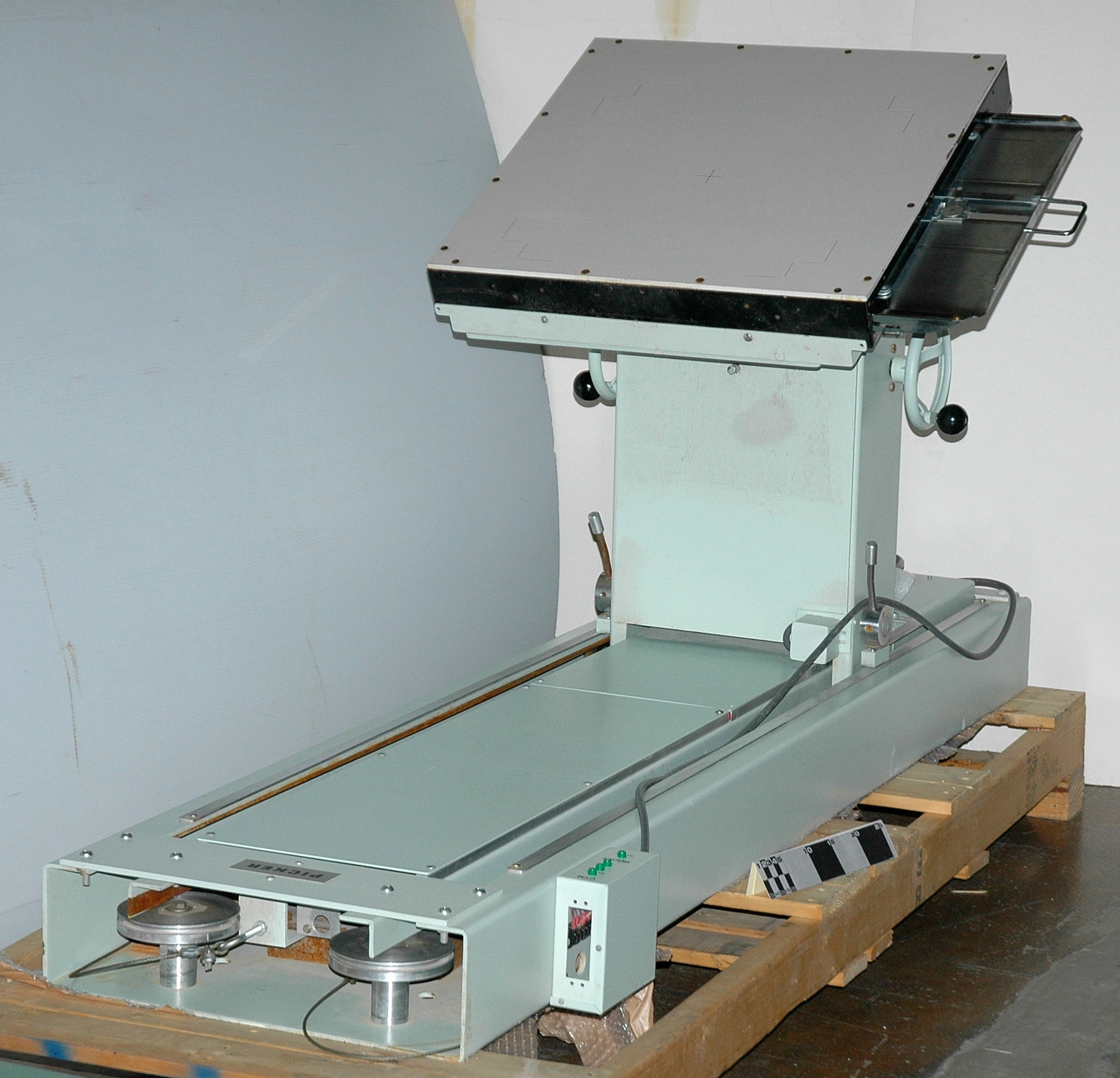
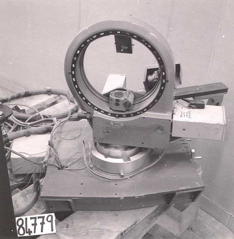
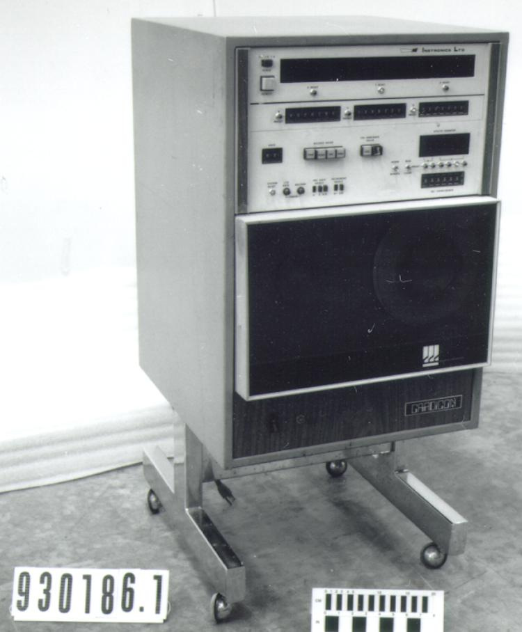
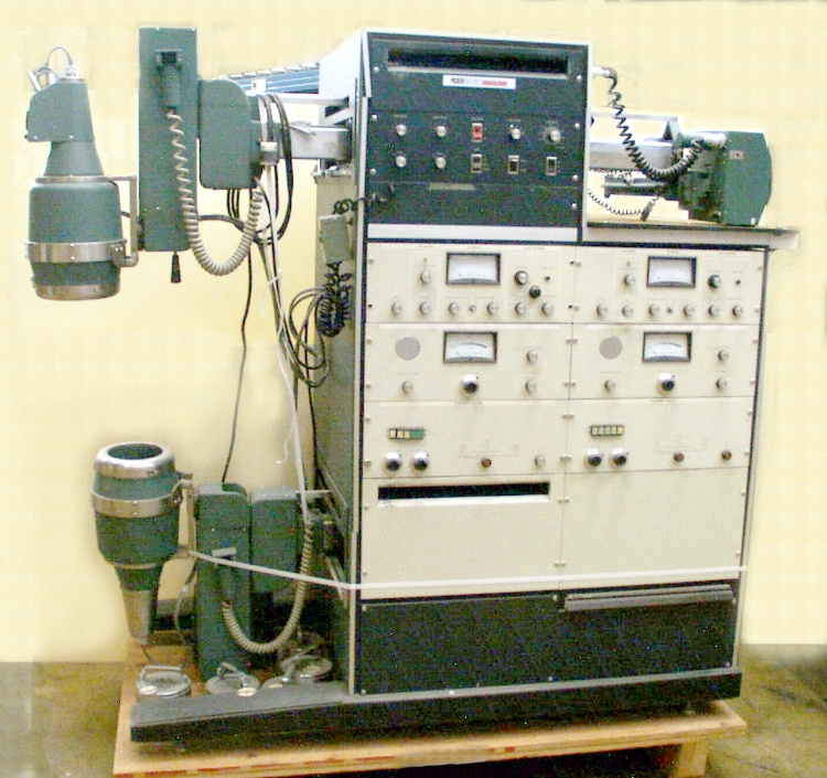
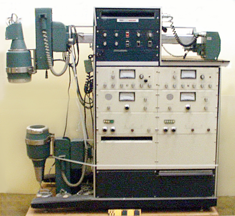
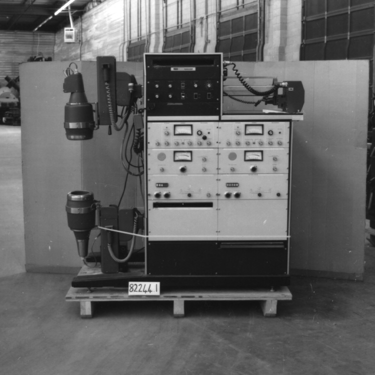
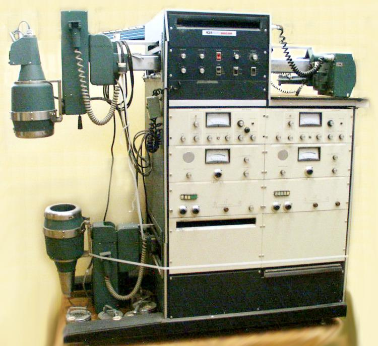
Scanner, radioisotope
Use this image
Can I reuse this image without permission? Yes
Object images on the Ingenium Collection’s portal have the following Creative Commons license:
Copyright Ingenium / CC BY-NC-ND (Attribution-NonCommercial 4.0 International (CC BY-NC 4.0)
ATTRIBUTE THIS IMAGE
Ingenium,
1982.0244.001
Permalink:
Ingenium is releasing this image under the Creative Commons licensing framework, and encourages downloading and reuse for non-commercial purposes. Please acknowledge Ingenium and cite the artifact number.
DOWNLOAD IMAGEPURCHASE THIS IMAGE
This image is free for non-commercial use.
For commercial use, please consult our Reproduction Fees and contact us to purchase the image.
- OBJECT TYPE
- N/A
- DATE
- 1971
- ARTIFACT NUMBER
- 1982.0244.001
- MANUFACTURER
- Picker X-ray Mfg.
- MODEL
- DUAL MAGNASCANNER/2806N
- LOCATION
- Cleveland, Ohio, United States of America
More Information
General Information
- Serial #
- 205
- Part Number
- 1
- Total Parts
- 16
- AKA
- N/A
- Patents
- N/A
- General Description
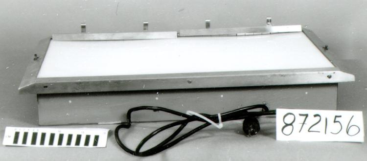
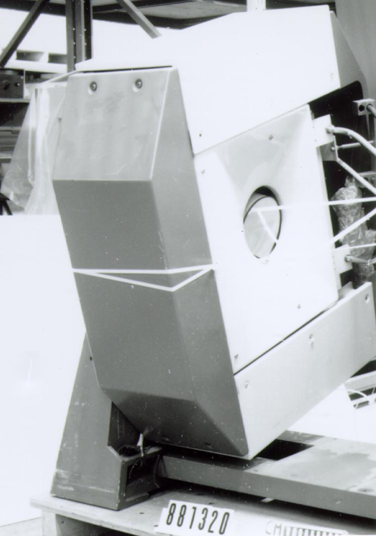
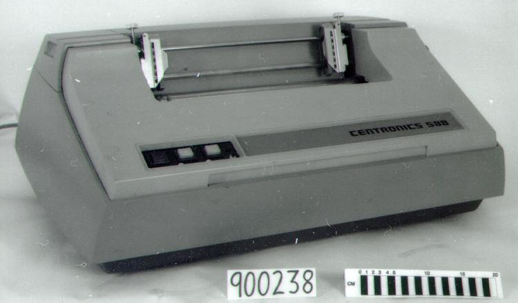
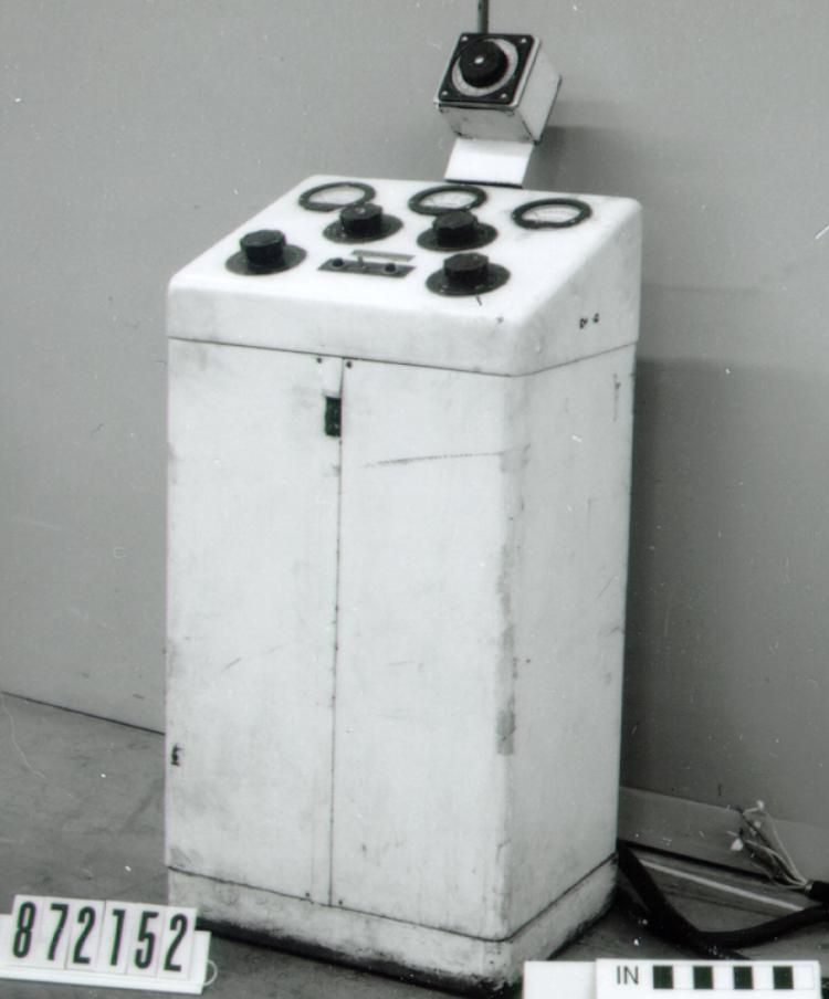
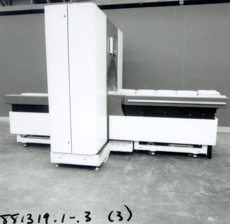
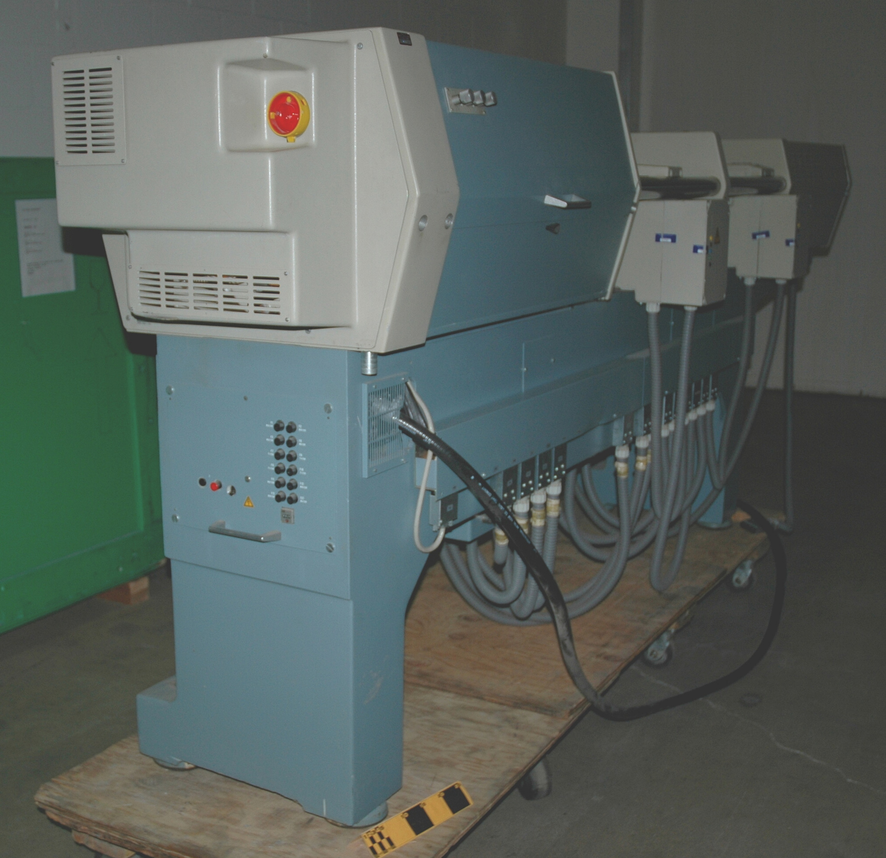
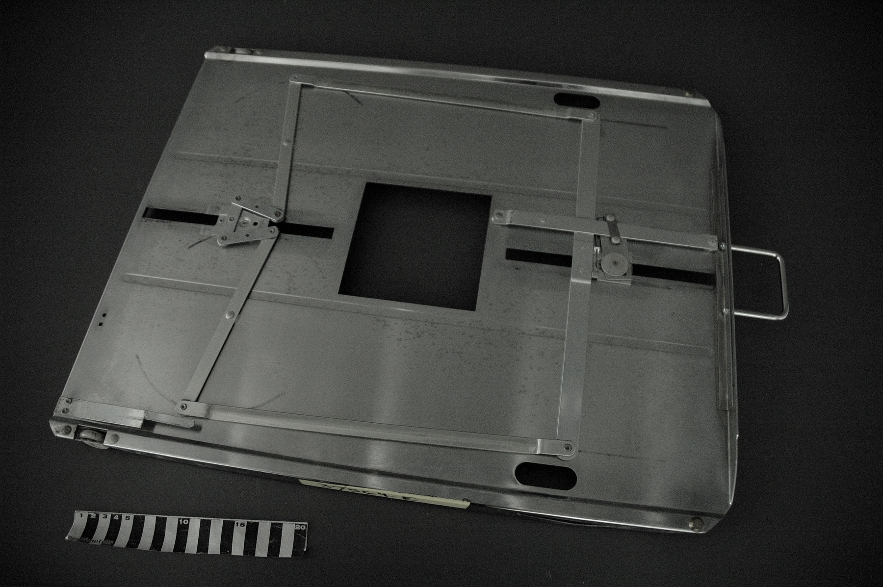
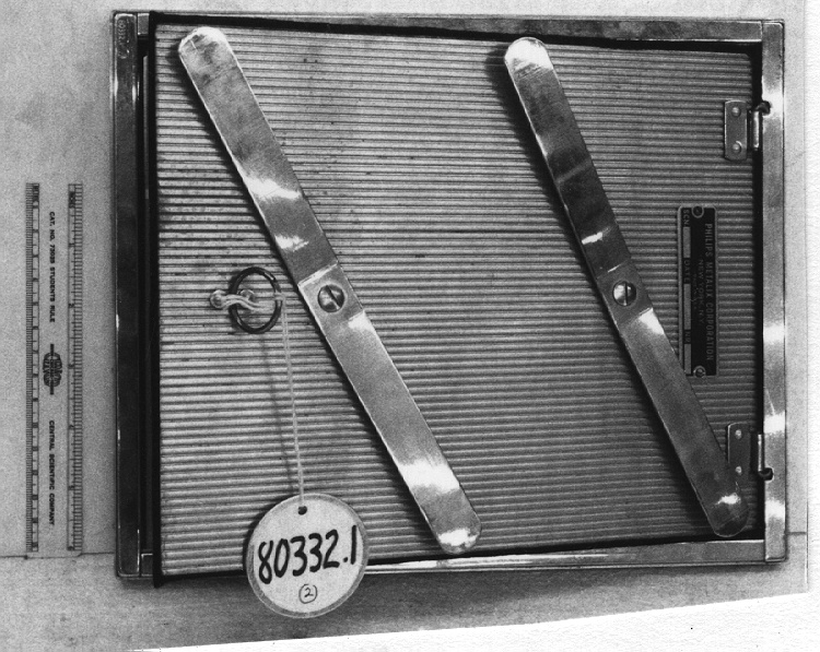
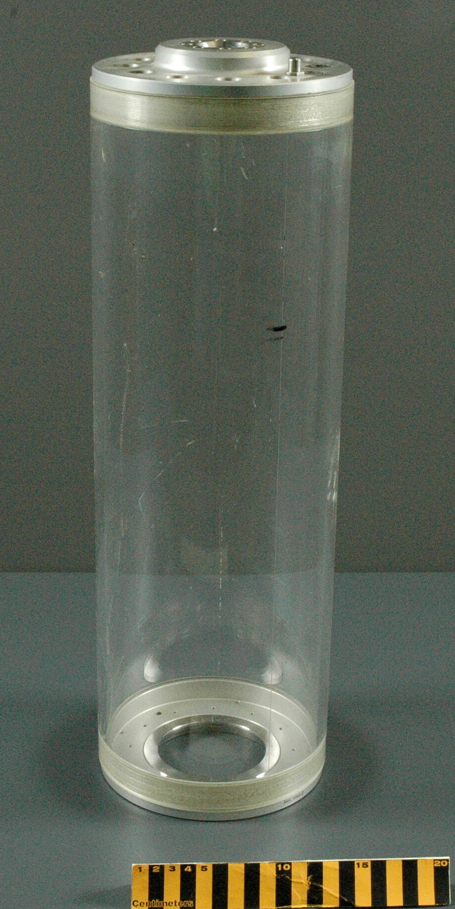
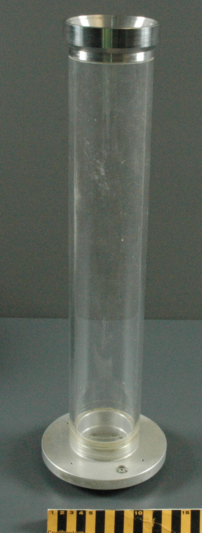
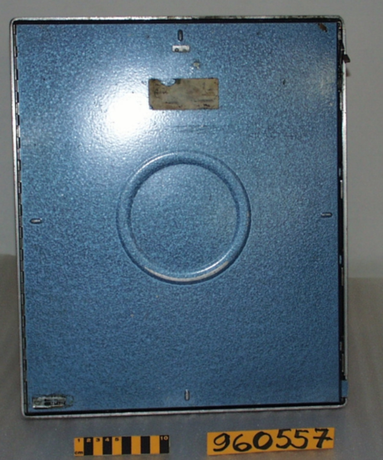
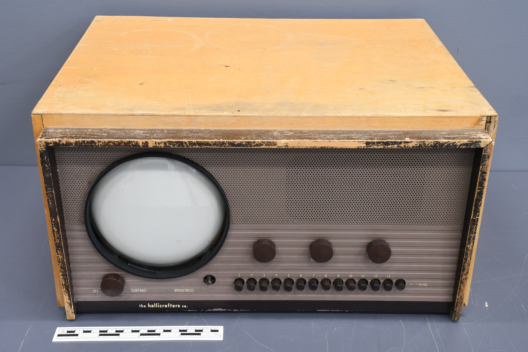
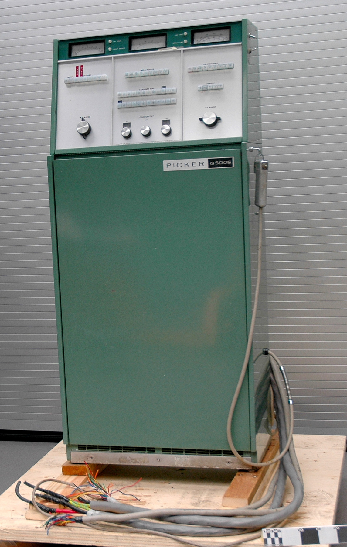
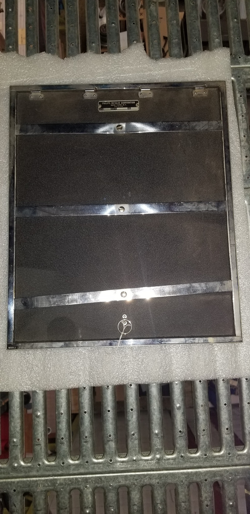
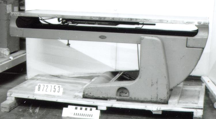
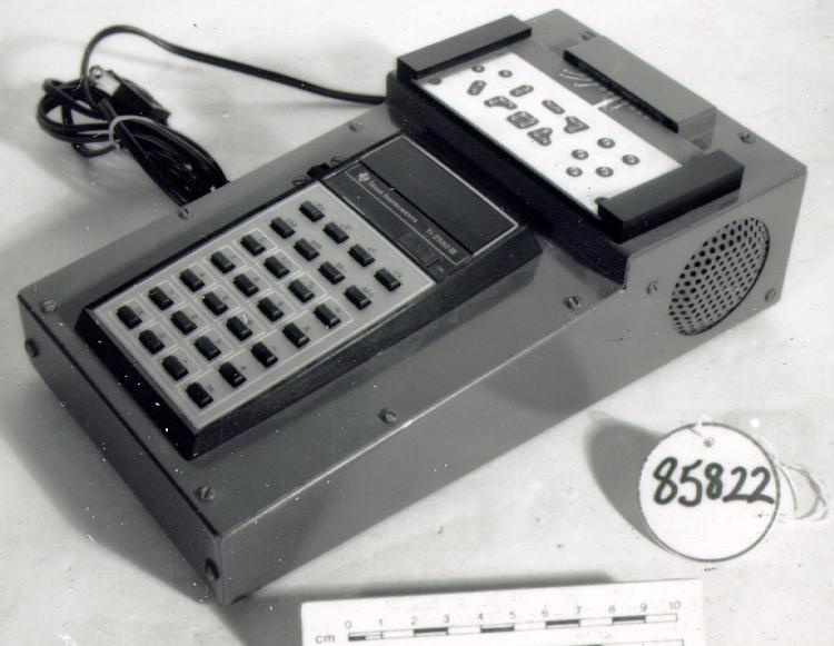
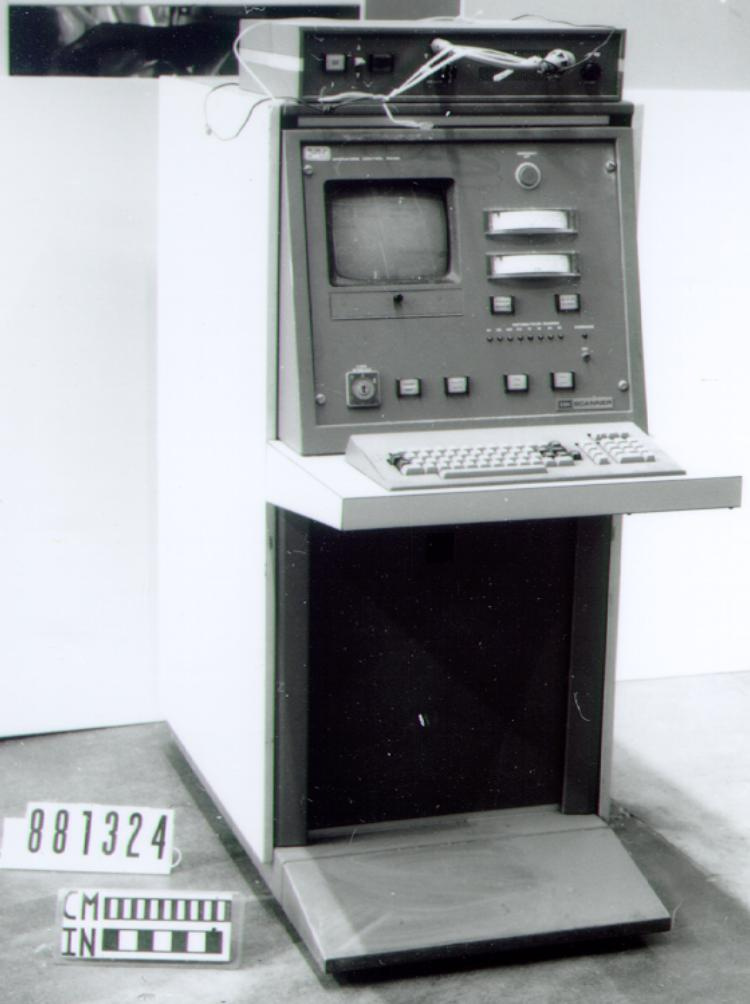
- METAL CASING & COMPONENT PARTS; SYNTHETIC PANELS, KNOBS, BUTTONS, SWITCHES, PRINTER TABLE, CORDS, ETC.; GLASS WINDOWS
Dimensions
Note: These reflect the general size for storage and are not necessarily representative of the object's true dimensions.
- Length
- 175.0 cm
- Width
- 81.0 cm
- Height
- 171.0 cm
- Thickness
- N/A
- Weight
- 684.9
- Diameter
- N/A
- Volume
- N/A
Lexicon
- Group
- Medical Technology
- Category
- Radiology
- Sub-Category
- N/A
Manufacturer
- AKA
- Picker
- Country
- United States of America
- State/Province
- Ohio
- City
- Cleveland
Context
- Country
- Canada
- State/Province
- Ontario
- Period
- PURCHASED NEW FOR $20,000 IN APRIL 1971, AND USED UNTIL JULY 1982.
- Canada
-
Unknown - Function
-
TO PLOT OR MAP THE INTENSITY DISTRIBUTION OF A RADIOACTIVE ISOTOPE IN A LOCALIZED AREA OF THE BODY, IN ORDER TO ASSIST IN DIAGNOSIS OF DISEASE. - Technical
-
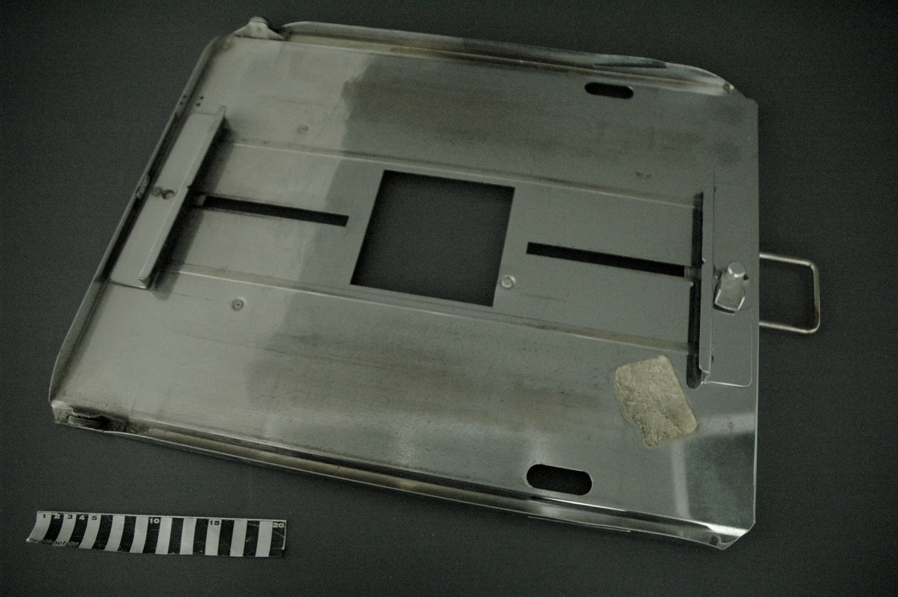
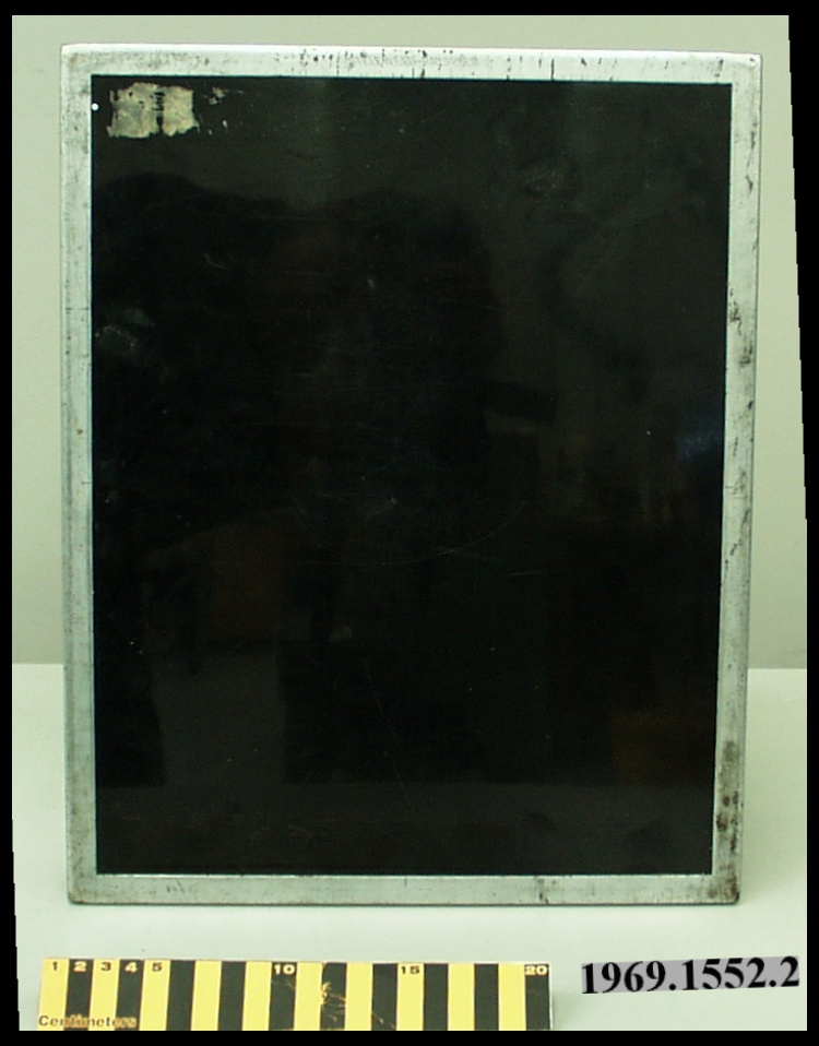
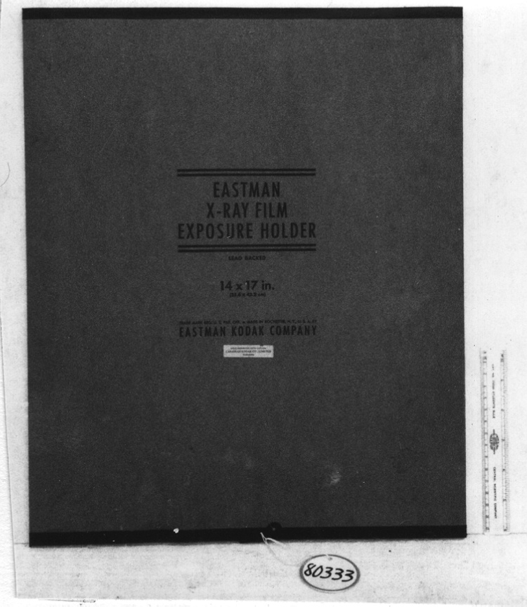
AT THE TIME OF ITS PURCHASE (1971), THIS SCANNER WAS THE MOST ADVANCED DEVICE OF ITS TYPE. IN OPERATION, RADIATION FROM THE SOURCE IMPINGES ON THE DETECTING CRYSTAL IN A SCINTILLATION PROBE (THE RADIATION FIRST PASSES THROUGH THE COLLIMATOR WHICH PERMITS THE CRYSTAL TO VIEW ONLY THE RADIATION DIRECTLY BELOW IT DURING THE SCAN). THE RADIATION ABSORBED BY THE CRYSTAL IS CONVERTED TO A LIGHT FLASH WHICH, THROUGH A SERIES OF STEPS, IS CONVERTED TO AN IMAGE ON EITHER ELECTROSENSITIVE PAPER (DOT RECORDING) OR X-RAY FILM (PHOTO RECORDING) ON 14 X 17 INCH FILM. THE RESULTING MAP CONSISTS OF MULTIPLE BLACK DOTS ON A WHITE OR CLEAR GROUND, THE FREQUENCY OF DOT OCCURRENCE INDICATING THE DEGREE OF CONCENTRATION OF RADIOACTIVE MATERIAL & HENCE THE LEVEL OF ACTIVITY OF THE ORGAN. THIS EXAMPLE HAS THE COLOUR DOT RECORDING OPTION AS WELL. (MANUALS T55-627 & T55- 564.) - Area Notes
-
Unknown
Details
- Markings
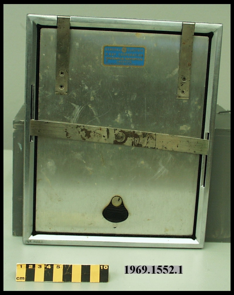
- BLACK & SILVER MFR'S PLATES ON SCANNER, PROBE, COLOUR PRINTER, & COLLIMATORS/ BLACK, WHITE, RED, & BLUE LETTERING & MARKING ON CONTROL PANELS/ INCISED LETTERING ON CASSETTES READING 'Picker, Made in U.S.A.'/ '265' INCISED ON UNDERSIDE OF.6 COLLIMATOR
- Missing
- UNKNOWN.
- Finish
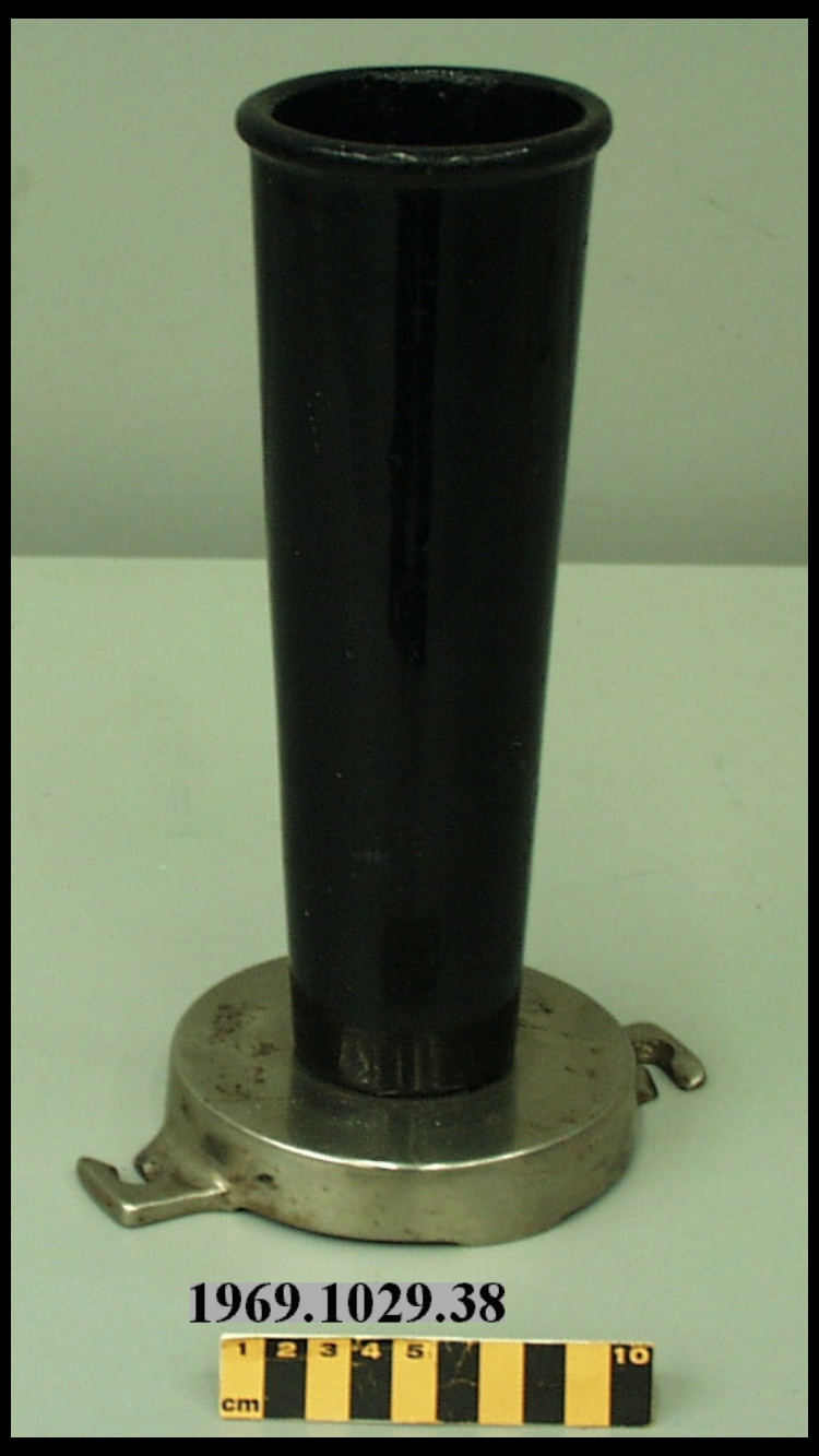
- METAL CASING PAINTED MEDIUM GREEN. FRONT PANELS PAINTED DARK GREEN & BEIGE. PLATED TRIM. BLACK SYNTHETIC & METALLIC KNOBS. MULTICOLOURED SYNTHETIC BUTTONS. BLACK SYNTHETIC PRINTER TABLE. PLATED COLLIMATORS. BLACK PAINTED & PLATED CASSETTES.
- Decoration
- N/A
CITE THIS OBJECT
If you choose to share our information about this collection object, please cite:
Picker X-ray Mfg., Scanner, radioisotope, circa 1971, Artifact no. 1982.0244, Ingenium – Canada’s Museums of Science and Innovation, http://collection.ingeniumcanada.org/en/id/1982.0244.001/
FEEDBACK
Submit a question or comment about this artifact.
More Like This
