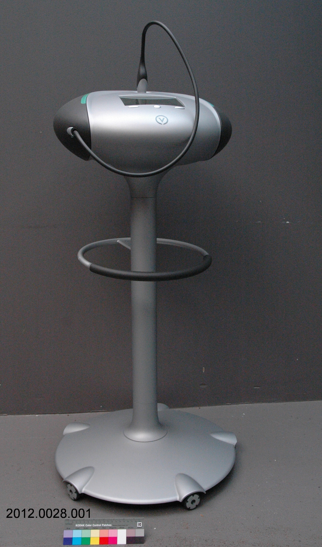
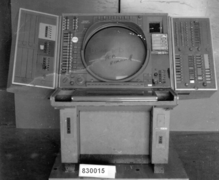
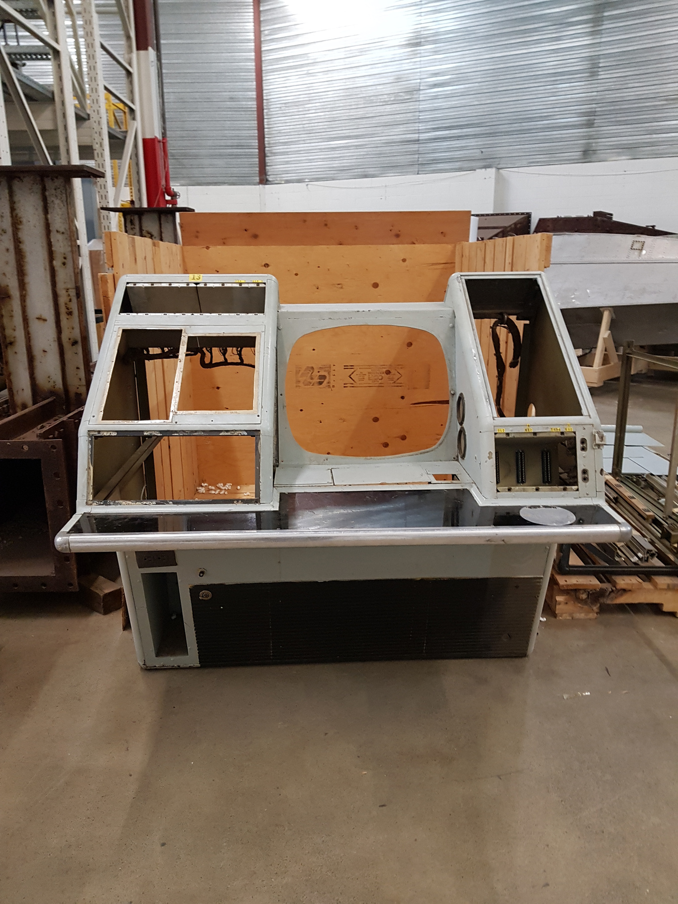
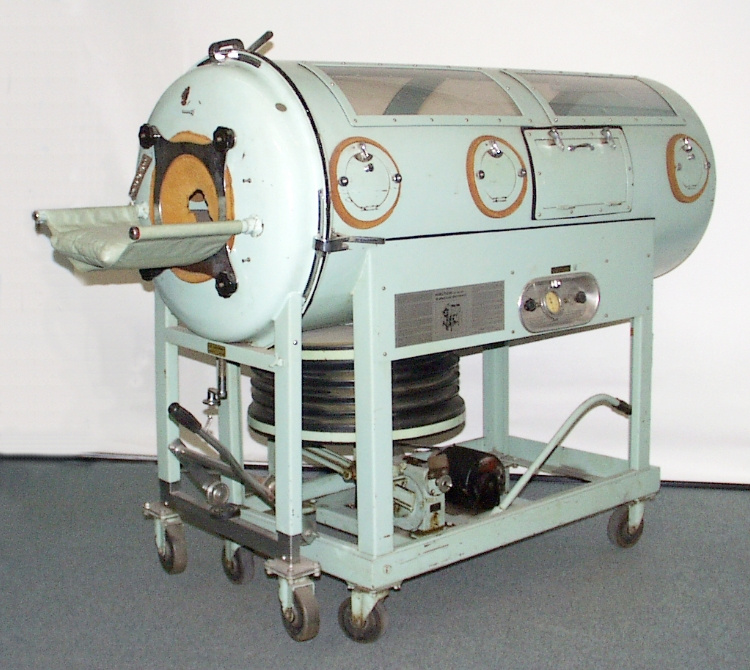
Imaging system model
Use this image
Can I reuse this image without permission? Yes
Object images on the Ingenium Collection’s portal have the following Creative Commons license:
Copyright Ingenium / CC BY-NC-ND (Attribution-NonCommercial 4.0 International (CC BY-NC 4.0)
ATTRIBUTE THIS IMAGE
Ingenium,
2012.0028.001
Permalink:
Ingenium is releasing this image under the Creative Commons licensing framework, and encourages downloading and reuse for non-commercial purposes. Please acknowledge Ingenium and cite the artifact number.
DOWNLOAD IMAGEPURCHASE THIS IMAGE
This image is free for non-commercial use.
For commercial use, please consult our Reproduction Fees and contact us to purchase the image.
- OBJECT TYPE
- display/full scale/static
- DATE
- 2010
- ARTIFACT NUMBER
- 2012.0028.001
- MANUFACTURER
- ProtoTechnologies Inc.
- MODEL
- Verisante Aura
- LOCATION
- Liberty Lake, Washington, United States of America
More Information
General Information
- Serial #
- N/A
- Part Number
- 1
- Total Parts
- 2
- AKA
- N/A
- Patents
- N/A
- General Description
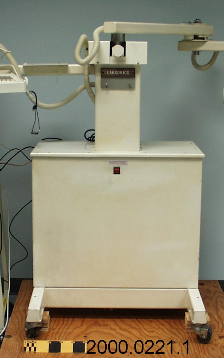
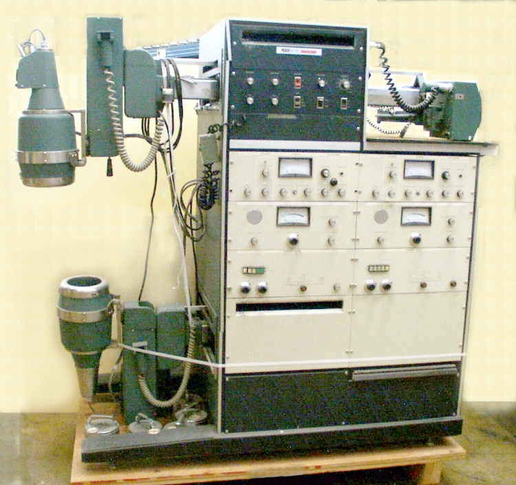
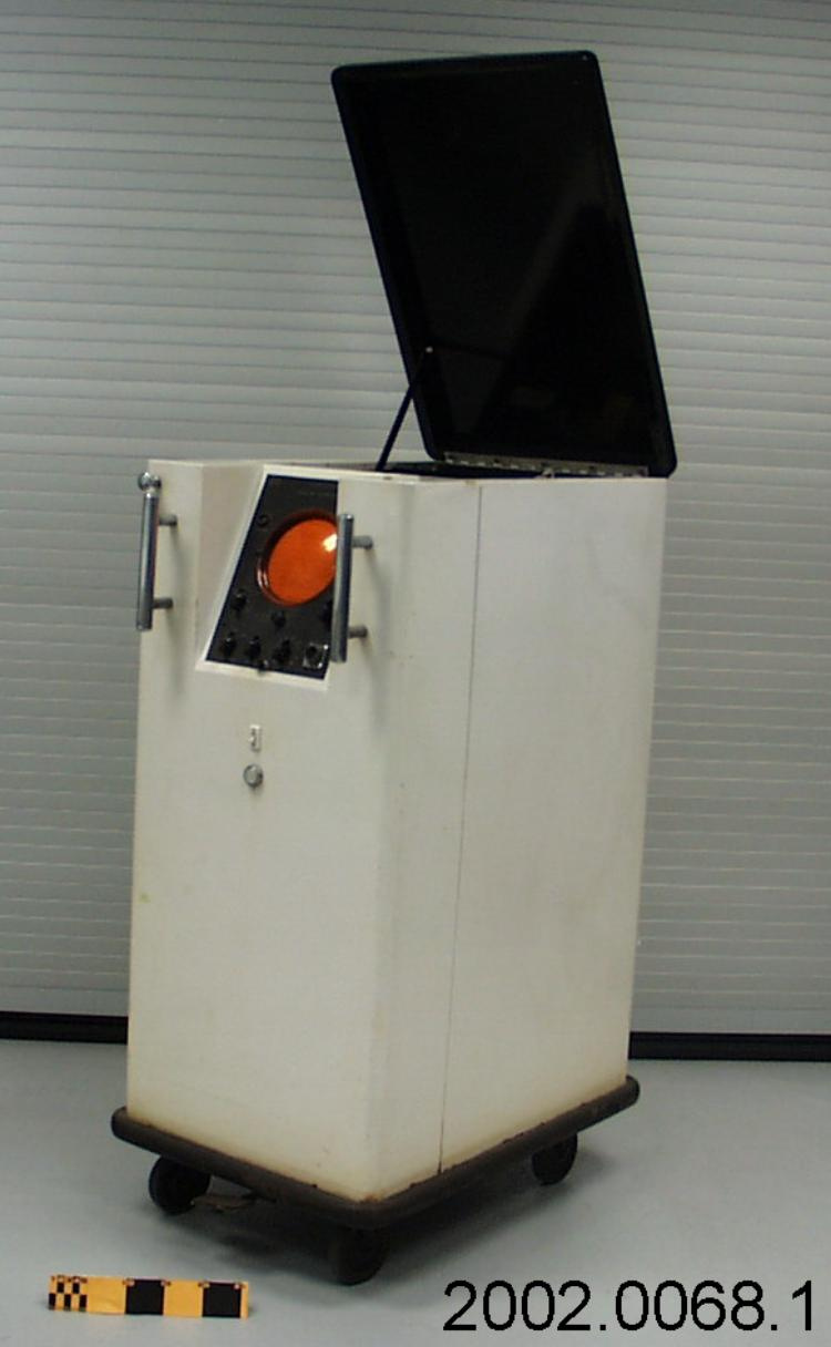
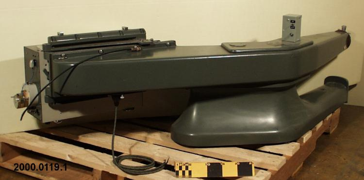
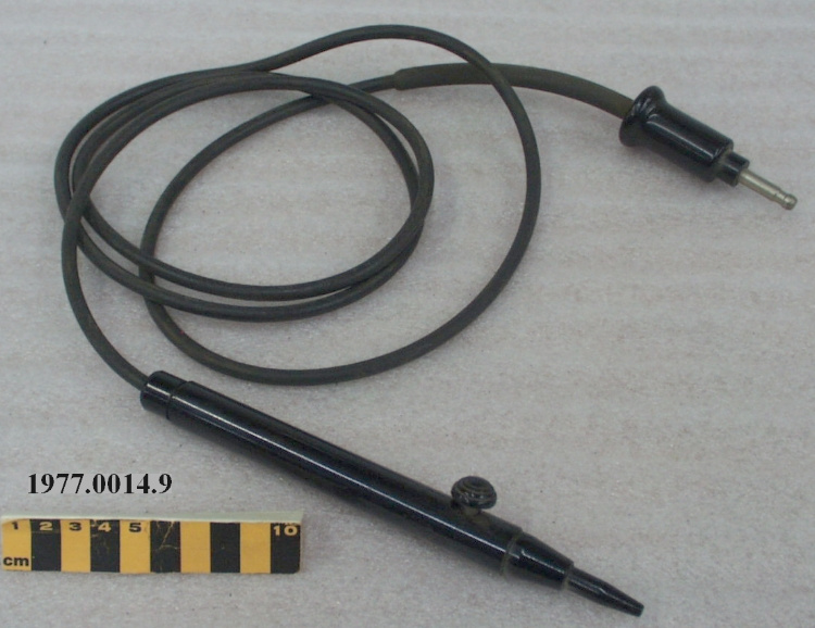
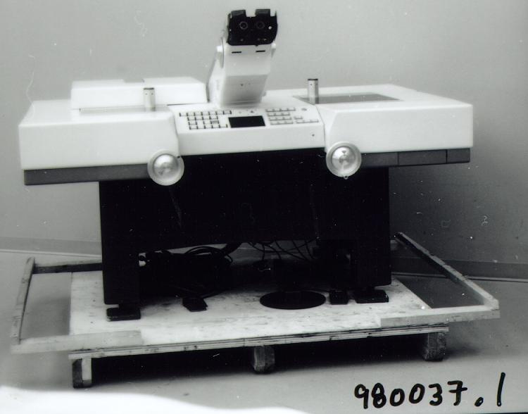
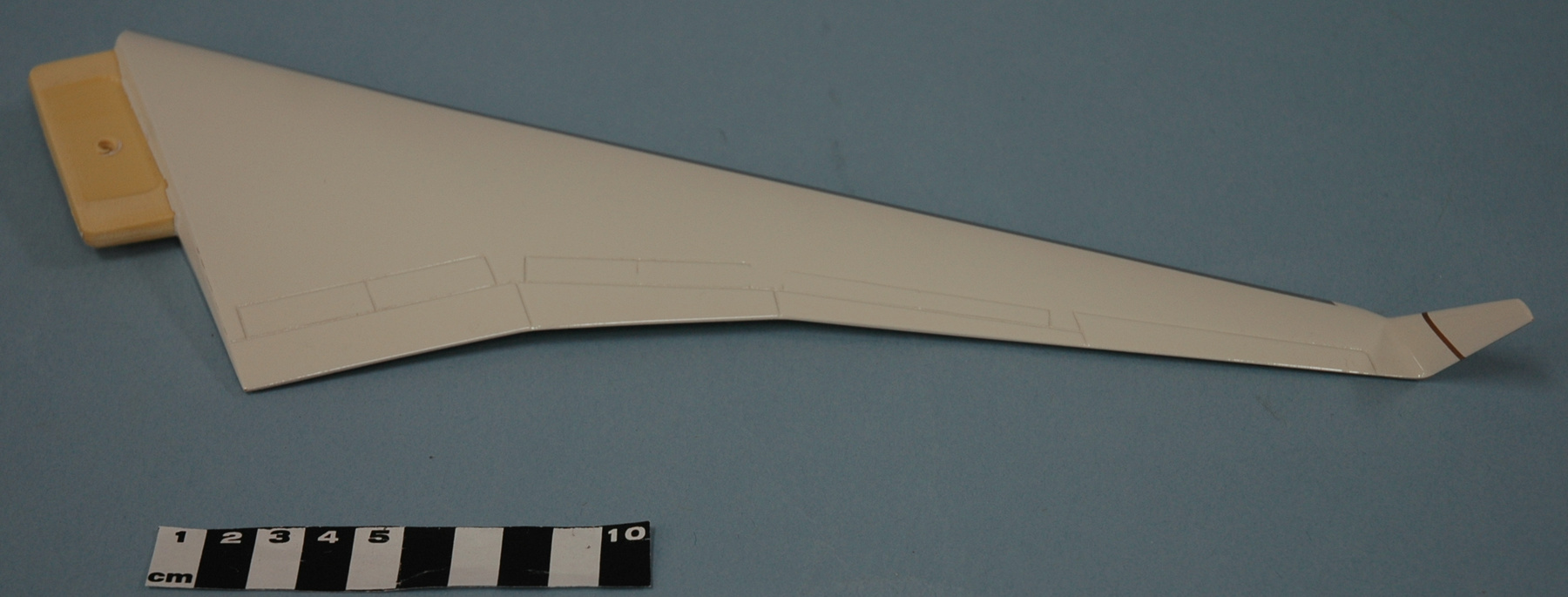
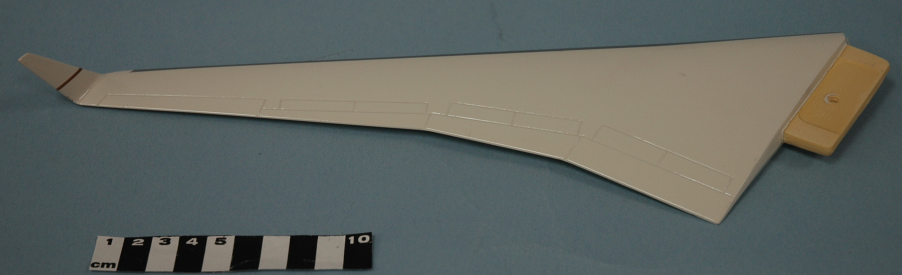
- Epoxy resin model is predominantly silver grey: features darker grey trim on bulbous display console, on portion of ring surrounding central support post, and on cord and wand [attachment]; clear synthetic display screen; green band trims console face; bright red on-off switch mock-up [?]; grey synthetic wheels/castors.
Dimensions
Note: These reflect the general size for storage and are not necessarily representative of the object's true dimensions.
- Length
- N/A
- Width
- N/A
- Height
- 131.0 cm
- Thickness
- N/A
- Weight
- N/A
- Diameter
- 58.0 cm
- Volume
- N/A
Lexicon
- Group
- Medical Technology
- Category
- Medical equipment
- Sub-Category
- N/A
Manufacturer
- AKA
- Proto
- Country
- United States of America
- State/Province
- Washington
- City
- Liberty Lake
Context
- Country
- United States of America
- State/Province
- California
- Period
- Model displayed in 2011.
- Canada
-
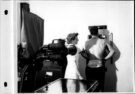
The emergence of Verisante Aura has deep roots in Canadian medical and scientific fields dating to the earliest days of the x-ray . British Columbia and the BC Cancer agency, have a long history of developing new technologies for cancer detection . The Triumf facility at UBC also has a unique tradition of combining physics and medicine. Verisante brings together both of these traditions. Medical imaging has always been at the forefront of cancer detection in Canada, e.g. X-rays, MRI and CAT technologies have been used to detect numerous forms of cancer. The Verisante Aura uses an optical (non-radioactive technique) to scan skin surface for indicators of cancer. In the form of early detection, it replaces biopsy and the trained eye of the dermatologist. Cancer detection technologies have also played an important role in public health and medical policy in Canada. New forms of diagnosis such as x-ray fluoroscopy and mammography have been a key to the development of groundbreaking provincial Medicare programs. In 1930 the government of Saskatchewan introduced the first full cancer coverage in the country based on diagnostic and treatment technologies. In the present medical context, the early detection of skin cancer is seen as a key element for preventing death due to melanoma and saving health care costs. [Ref. 1] - Function
-
3-D marketing model created and used for promotional purposes. - Technical
-
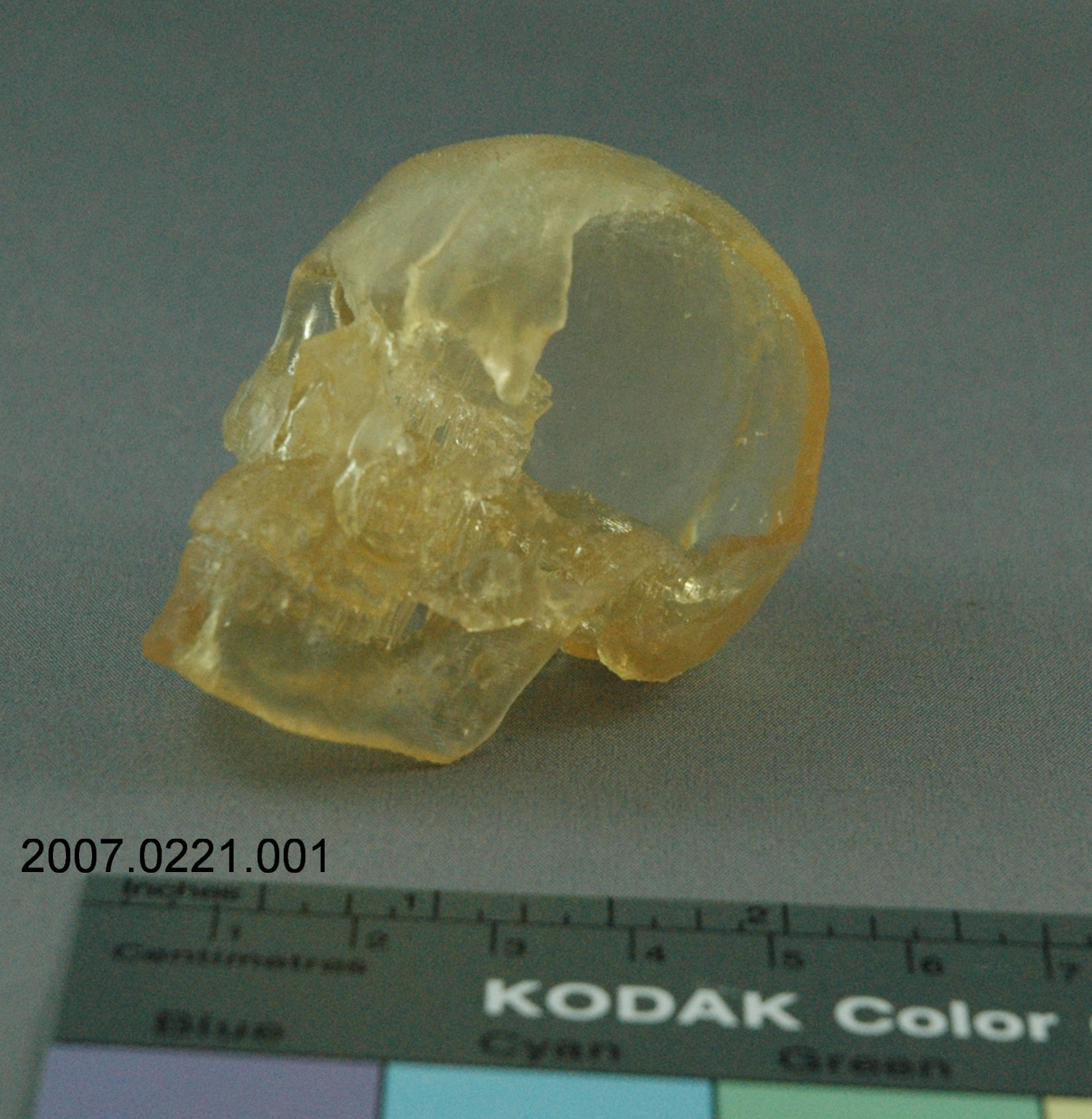
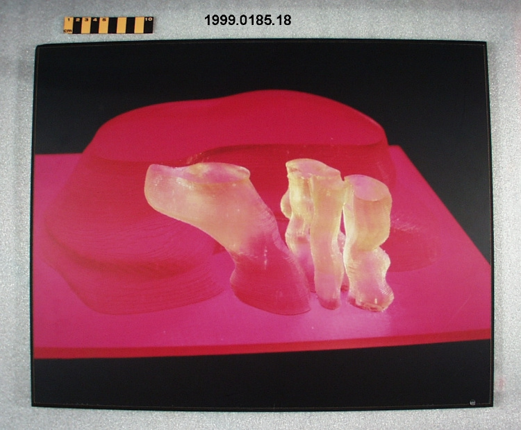
The technical significance of Verisante technology is in the use and application of Raman spectroscopy to the detection of skin cancer. According to Verisante, they are the first in the world to apply Raman spectroscopy to the detection of skin cancer. The model represents the prototype Verisante Aura is manufacturing – a real-time non-invasive imaging technology which allows better targeting of tumors and less risk to patients. The technology uses Raman spectroscopy to detect early forms of skin cancer, and is marketed as an “automatic process of diagnosis allowing rapid scanning of at-risk individuals, reducing patient wait times and costs, and improving patient outcomes and comfort”. The model is made of epoxy resin from a 3-D prototyping technique called stereolithography (SLA). Stereolithography (SLA) is prototyping technique introduced in the early 1980’s. A laser which generates a small intense beam of ultraviolet (UV) energy is moved by the machine’s optical scanning system across a vat of photosensitive liquid polymer, building a part or device layer by layer. As the UV beam comes in contact with the polymer, it solidifies to create the finished product. [Ref. 1] - Area Notes
-
Unknown
Details
- Markings
- Logo appears on console housing, on wand and on ring surrounding central support post.
- Missing
- Mock-up is complete in itself.
- Finish
- Epoxy resin model is predominantly silver grey: features darker grey trim on bulbous display console, on portion of ring surrounding central support post, and on cord and wand [attachment]; clear synthetic display screen; green band trims console face; bright red on-off switch mock-up [?]; grey synthetic wheels/castors.
- Decoration
- Stylized "V' within circular border forms company logo.
CITE THIS OBJECT
If you choose to share our information about this collection object, please cite:
ProtoTechnologies Inc., Imaging system model, 2010, Artifact no. 2012.0028, Ingenium – Canada’s Museums of Science and Innovation, http://collection.ingeniumcanada.org/en/id/2012.0028.001/
FEEDBACK
Submit a question or comment about this artifact.
More Like This


2012.0028.001